Vp Shunt Procedure Steps
The surgery will take about 1 hour. Scalp incision and burr hole positioning.
Ventriculoperitoneal Shunt India Reviews Is It Effective
Semilunar incision usually on the right side virgin patients Avoid coagulating the skin Create a subcutaneous pocket to house the reservoir Occipital.

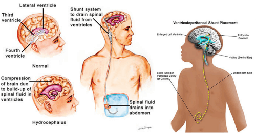
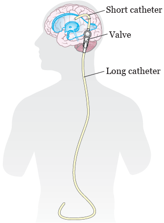
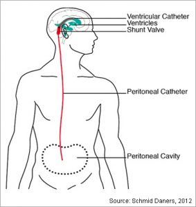
Vp shunt procedure steps. Usually VP shunts are placed to treat hydrocephalus hydro water. It will be moved into the correct. Ventriculoperitoneal Shunt Introduction Ventriculoperitoneal VP shunt insertion is an operation to place a catheter into a brain ventricle to drain cerebrospinal fluid CSF from the ventricular system. The following technique is used. It is moved under your skin on your neck down to your chest and then usually into the stomach area ventriculo-peritoneal shunt. During a VP shunt procedure a small catheter or thin flexible tube is implanted in the brain usually behind the ear.
6 Steps for the Perfect VP Shunt. Your VP shunt surgery will take place in the operating room while youre asleep. However VPL shunts are exposed to a negative subatmospheric intrathoracic pressure with each breathing cycle which imparts an active sucking force. For a few weeks after surgery you may have headaches. The surgery will take about 1 hour. The tube is then run under the skin to the abdominal cavity allowing excess fluid to drain and be reabsorbed into the bloodstream.
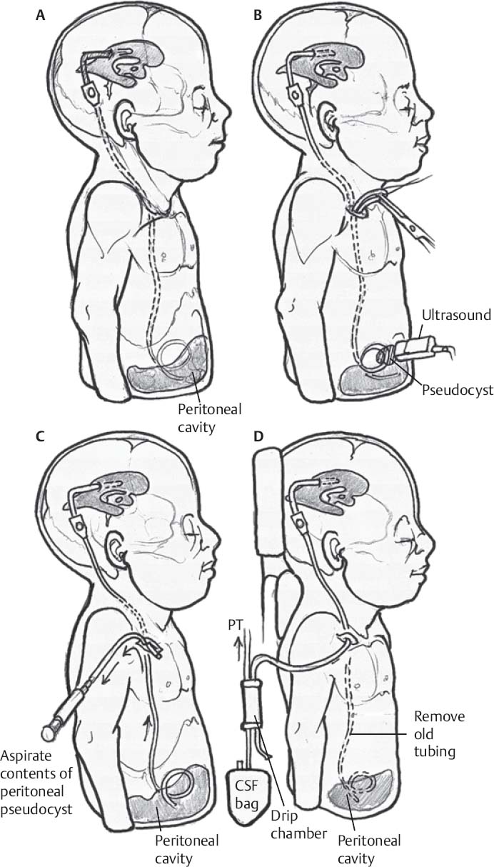
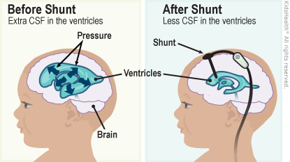
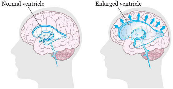
A ventriculoperitoneal VP shunt is a medical device that relieves pressure on the brain caused by fluid accumulation. Once youre asleep the doctor will shave off some hair near the area where theyll make the incision surgical cut on your head. A A 25-gauge needle is placed in the valve component of a Cordis Unishunt ventriculoperitoneal shunt closed arrow. We describe a minimally invasive laparoscopic technique. If the ventricular catheter is placed frontally a length of distal shunt tubing is secured to the outflow port of the unidirectional shunt. Place the top end of the shunt into the ventricles of your brain.
2-3 cm from midline 3-4 cm above the inion Frontal. Another catheter will then be inserted through the hole under the skin behind your ear. Your VP shunt surgery will take place in the operating room while youre asleep. During VP shunt surgery the doctor placed two small tubes catheters and a valve under your skin. A 1-way shunt valve should be chosen. A small pump or valve may also be attached to the catheter and implanted behind the ear.
After surgery your neck or belly may feel tender. 2-3 cm from midline just in front of the coronal suture or at the fontanel angle neonates Dandy-Walker. Through the incision the vp shunt surgeon will drill a hold right into the skull in order to reach the ventricles of the brain. A small catheter is used for draining the excessive fluid accumulated. If playback doesnt begin shortly try restarting your device. This force can lead to shunt overdrainage.
Videos you watch may be added to the TVs watch history and influence TV recommendations. Once youre asleep the doctor will shave off some hair near the area where they will make the incision surgical. A shunt reservoir is seen just proximal to the valve open arrow. This fluid will then drain into the peritoneal space abdominal cavity. You will probably feel tired but you should not have much pain. Ventriculoperitoneal shunting VPS remains one of the alternative choices for the surgical treatment of hydrocephalus.
A shunt reservoir is again seen proximal to the valve open arrow. The distal resistance of VP and ventriculoatrial shunts is assumed to be close to zero in most patients. Clean the skin with an antiseptic Apply a sterile fenestrated drape over the incision site Insert a small 23 ga butterfly needle perpendicular to the skin into the reservoir Evaluate for spontaneous CSF filling the. VP shunting is a surgical procedure that primarily treats a condition called. During the last two decades laparoscopy has been utilized to facilitate the placement of the abdominal portion of the shunt. B Contrast material is injected into the valve closed arrow.
The second catheter is made to pass subcutaneously through the chest and the abdominal region.
Lumboperitoneal Shunt Fort Worth Brain And Spine Institute

Stages Of The Subgaleal Shunt Technique Download Scientific Diagram

Illustrations Showing The Surgical Steps Involved In This Technique A Download Scientific Diagram

Accurate Placement Of Parieto Occipital Shunt Ventricular Catheter Use Of Craniometrics And Technical Note Springerlink

Scholarworks Gsu Edu
Illustrations Showing The Surgical Steps Involved In This Technique A Download Scientific Diagram

Using Laparoscopy For Ventriculoperitoneal Sh Eurekalert
Jew Eat Yet Due Dates And Vp Shunts
Shunt Externalization Neupsy Key
Laparoscopic Ventriculoperitoneal Shunt Repositioning A Novel Technique Sages Abstract Archives
About Your Ventriculoperitoneal Vp Shunt Surgery Memorial Sloan Kettering Cancer Center
Vp Shunts Dayton Children S
Ventriculoperitonial Shunt Pacific Adult Hydrocephalus Center
About Your Ventriculoperitoneal Vp Shunt Surgery Memorial Sloan Kettering Cancer Center

Posting Komentar untuk "Vp Shunt Procedure Steps"