Normal Distance Between L5 And S1
The nerve supply that reaches the glutes are called inferior gluteal nerves L5 S12. The ventral rami divide into anterior and posterior divisions that join to form the terminal nerves.
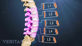
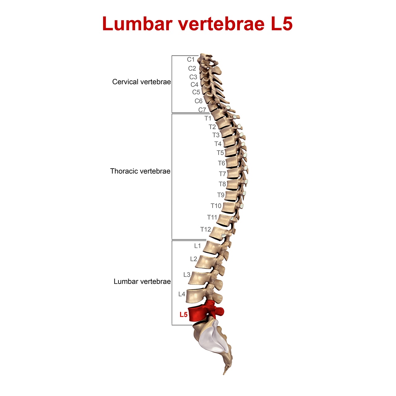
Lumbar Spinal Nerves
Contraction of EAS around examiners finger.

Normal distance between l5 and s1. See appendix 3-4 and see color plates. The plexus is located in the posterior abdominal wall between the psoas major and quadratus lumborum muscles see Figure 15. As much as 75 of lumbar lordosis occurs between L4 and S1 with 47 occurring at L5S1 normal alignment the vertical axis runs from the center of C2 to the anterior border of T7 to the middle of the T12L1 disc posterior to the L3 vertebral body and crosses the posterior superior corner of the sacrum. Venous plexus in posterior pelvis accounts for 90 of the hemorrhage associated with pelvic ring injuries. FIGURES 4A and 4B. He was concerned for one of his patients who developed a sudden foot drop without lower back pain.
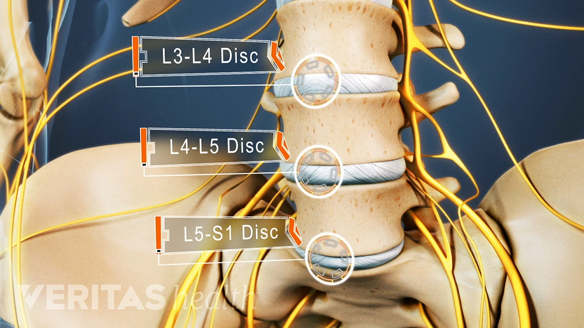
In an L4-5-level herniation the L5 spinal nerve is usually involved. L5 CNAV messages begin and end at startend of GPS week plus an integer multiple of 6 seconds this applies to the beginning of the first bit to contain information about a message as is the case for L2 CNAV. 2 for L34 L5 and S1. With its normal 3 curves keeps the spinal muscles active so they can share the load placed on the. A 2016 study published in the Journal of Neurosurgery found that over 90 percent of low-back disc herniations involved the L4-5 or L5-S1 levels the two lowest vertebral levels in the spine. In patients with a lumbardized S1 the last fully mobile level is usually L6-S2 and the functional L5 nerve root corresponds with the L6 nerve root.
Compressed L5 nerve blue arrow within the lateral recess. This is called an extrusion because the distance between the edges of the disc material is greater than the distance at the base. Anterolisthesis most commonly occurs at the L5-S1 level with anterior translation. Although the MRI of her lower back was positive for L4-5 L5-S1 Disc bulges with nerve root impingement the neurologist was convinced that there was a nerve impingement at the knee and the foot drop was not coming from the back. Muscles are responsible for locomotion and play an important part in performing vital body functions. Often accompanied by foot drop Gluteus medius and minimus weakness or damage eg.
Anterolisthesis most commonly occurs in the lower back lower lumbar spine but can also occur in the cervical spine and rarely except for trauma in the thoracic spine. Broadcast on the L5 frequency 117645 MHz 1023 MHz 115 which is an aeronautical navigation band. I had suffered from tremendous pain in the right leg prior to the operation most of that not gone but for Restless Legs Drop Foot and this tight sock feeling. Introduction edit edit source. Up to 10-15 of patients will sustain neurologic injury. The International Standards for Neurological.
At the level of the lateral recess there is a focal herniation of disc material compressing the L5 nerve yellow arrow. A systematic examination of dermatomes and myotomes thus would allow a clinician to determine the affected segments of the spinal cord. Decrease disc heights were seen in 31 disc levels from which decreased disc height common at L5-S1 level 10ie. All other ROM measurements within functional limits no pain. The most common location for disc injury is at L4-5 and L5-S1. Under normal circumstances the discs act to transfer and absorb loads.
Hip ROM 75 of normal no increase in pain with movement. Graded as present or absent. I had a Laminectomy in August L3L4 L4L5 L5S1. Muscle musl a bundle of long slender cells muscle fibers that have the power to contract and hence to produce movement. Another insertion point is the iliotibial tract connecting to the tibia. In general the front part of the disc is the strongest and the back and sides are weaker and susceptible to herniation and other disc disorders.
S1 represents the sacrum and is identified as the region of the spine that connects the spine to the pelvis. Mild spondylolisthesis is also evident at L5-S1. Anything including pain that prevents a person from working is a disability. The researchers concluded that patients with hip and lower back pain should be evaluated for leg length discrepancy. 22 Chang et al. 1 Research indicates that 90 of lumbar disc herniations occur at the L4-L5 or the L5-S1 disc space.
Two patients 183 showed changes of discitis. Layer signatures between ages were compared by identifying the top 10 most laminar genes evenly allocated between layers top 25 each in L23 L4 L5 and L6 and then calculating their. Can I qualify for SSDI. Spinal Cord Injury can severely impair or cease the conduction of sensory and motor signals as well as functions of the autonomic nervous system. Between the L2 and S3 levels the plexus is more complex. Agreed concluding that neurologic symptoms caused by the L6 nerve root compression resemble those of the L5 rather than the S1 nerve root compression in the normal configuration.
L5 and S1 are most common rectal exam to evaluate sphincter tone and perirectal sensation. Where it would be reasonably impracticable for constructional reasons to comply with the requirements of clauses 72431 and or 72432 and 72433 be as close as possible to the front andor rear respectively provided that where the distance between the front and rear lamps would then be less than 25 metres only the rearmost pair. They also protect the contents of the abdomen against injury and help support the body. The researchers found a strong connection between leg length discrepancy and degenerative disc disease at the L5-s1 spinal segment and the L4-L5 spinal segment. Low back pain risks increase when the compressive force at the L5-S1 lumbar 5 sacral 1 disc exceeds 770 lbs. Tears or tendonitis Poliomyelitis causing paralysis of the hip abductors.
L5 radiculopathy the superior gluteal nerve is formed from L4 L5 and S1 spinal roots. 3859 of the disc involvement. Read more about Causes of Lower Back Pain. 3226 of decreased disc height. Left Unenhanced T 1 -weighted axial magnetic resonance scan. I have this too on my right leg.
D vertical travel distance between the origin and the destination in inches F average frequency of lifts liftsminute. I have osteoarthritis on my back plus bone spurs Lumbar Spondylosis disk protrusion on L4 L5 disk protrusion on L5-S1 and degenerative changes on my lumbosacral spine. L5 Long Toe Extensors S1 Ankle Plantarflexors Motor Testing Test each of the ten key muscles Record numeric values only for research and test-taking purposes. Anon291136 September 12 2012. 45 on LE general exam. It is a horrible feeling I have to actually look sometimes to make sure I havent got a tight sock on.
The gluteus maximus attaches to the front of the legs by inserting into the gluteal tuberosity of the femur. L4 L5 disc involvement was common seen in 93 discs ie. The ventral rami join to form the terminal nerves. Lumbar ROM 75 of normal no increase in pain with movement. Mean distance of 62cm from the pubic symphysis.
Treating An L5 S1 Disc Herniation A Case Study Regenexx

A Lumbar Radiograph With Narrowing Of The L5 S1 Disc Space This Download Scientific Diagram
Where Is The L5 S1 Level Quora

Sagittal View Of L S Spine Showing Absolute Stenosis At L5 S1 Measuring Download Scientific Diagram

A Lumbar Radiograph With Narrowing Of The L5 S1 Disc Space This Download Scientific Diagram

After 2 Years From The L5 S1 Disc Replacement A Patient Complained For Download Scientific Diagram
Spine Radiology Key

Sagittal View Of L S Spine Showing Absolute Stenosis At L5 S1 Measuring Download Scientific Diagram
Anterior Fusion Of L5 S1 For Spondylolisthesis Type 1
Lumbar Discs

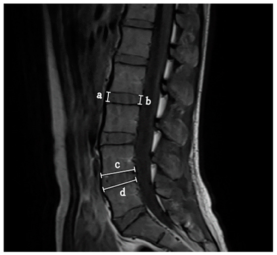
Measurement Of The Difference In Size Between L5 And S1 Is Shown In A Download Scientific Diagram
Ijerph Free Full Text Prediction Of Lumbar Disc Bulging And Protrusion By Anthropometric Factors And Disc Morphology Html
A Modic Change In Both Rostral And Caudal Endplates At L5 S1 Level Download Scientific Diagram

L5 S1 Disc Herniation With Hemilaminectomy And Discectomy Procedure Medical Exhibit Human Anatomy Drawing Disk Herniation Human Anatomy Drawing Medical Art
Posting Komentar untuk "Normal Distance Between L5 And S1"