Stress Fracture Mri With Or Without Contrast
Often the physician will do a study with and without contrast if there is fear of a malignancy as the study will offer the radiologist greater information about the mass. How Ultrasound And MRI Can Diagnose Calcaneal Bursitis.

Stress Fractures Diagnosis Treatment And Prevention American Family Physician
General pain MRI without contrast.

Stress fracture mri with or without contrast. When enough stress is placed on the bone it causes an imbalance between osteoclastic and osteblastic activity and a stress fracture may appear. Metatarsal stress fracture MRI without contrast Lis France ligament injury MRI without contrast FOReFOOTTOes ReGiON FROm mTP jOiNT TO disTal TiP Indication Preferred Study Trauma surgical hardware X-ray first. With x-ray flouro time Spine Pain Trauma Disc disease Radiculopathy Cord compression with no history of cancer No contrast Metastatic disease or spine tumor Infection multiple sclerosis. Upper Extremity Lower Extremity 73218 Upper 73718 Lower Abscess Myositis Ulcer Osteomyelitis. The most common patterns of a fatigue stress fracture on MRI are a linear uni-cortically-based abnormality of low-signal intensity surrounded by a larger ill-defined region of marrow edema or a linear cortical abnormality with adjacent muscular or soft tissue edema 7375. Radiographs are normal or equivocal.
If there is evidence of a stress fracture on an X-ray but your doctor needs to see the bones in more detail he or she may recommend a CT scan. Scintigraphy suspected to be a stress fracture. Often an MRI is the first diagnostic test used with or without an X-ray in assessing back pain and possibly diagnosing spondylolysis. Muscle fatigue can also play a role in the. MRI Abdomen With and Without Contrast MRI Pelvis With and Without Contrast 74183 72197 MRI Extremity NON JOINT. Bone marrow oedema is the earliest feature of acute osteomyelitis seen on MRI and can be detected as early as 1 to 2 days after the onset of infection.
Usually Appropriate MRI Ofoot without IV contrast. Most of the time shoulders do not need contrast. MRI is of growing importance for diagnosing bone pathology. MRI- With or without contrast. Application of additional gadolinium-DTPA contrast medium allows safe differentiation of stress fractures from pathological fractures due to the characteristic perifocal contrast enhancement in the T1 sequences. Call or Request an Appointment Online Today and Get Your MRI When its Convenient for You.
MRI Pelvis without contrast 72195 Fracture Trauma HipPelvis Pain MuscleTendon tear No Contrast ortho MRI Pelvis with and without contrast 72197 TumorMassCancerMets Septic arthritis. Bone for Fracture No MRI Pelvis Without Fibroids Yes MRI Pelvis With and Without Lumbar Plexis No MRI Pelvis Without. Callus formation indicates a more chronic stress fracture. Ad Get Your Magnetic Resonance Imaging at The Regions Most Preferred Orthopedic Practice. All patients with isolated edema in the femoral neck without a fracture line on the initial MRI had resolution with. In patients with a femoral neck stress fracture and fracture line the presence of a hip effusion on the initial MRI screening is an independent risk factor for fracture progression and early prophylactic surgical intervention should be considered.
This phenomenon is exemplified by a case report of a stress fracture of the. FractureStress Fracture MuscleTendon Tear MRI Non-Joint without Contrast Upper Extremity Lower Extremity 73218 73718 Extremity Non Joint. Upper Extremity Lower Extremity 73220 73720 Abscess Myositis Ulcer Osteomyelitis. Initial shoulder MR study detected no labral tear andor no tears of the tendons. Procedure Appropriateness Category Relative Radiation Level CT foot without IV contrast. Gadolinium contrast solution injected into the joint capsule Rule out labral tear of the hip Previous meniscectomy with a second knee injury.
Traumatic and atraumatic fractures are entities with distinct but often overlapping clinical manifestations imaging findings and management protocols. MRI Exams Contrast vs Non-Contrast Guide These suggestions are general guidelines that apply to the use of contrast for MRI exams provided. MRI pelvis wo contrast Coccyx fracture Pelvic fracture Pubic arthralgia Sacral fracture Sacroiliitis Sports hernia Stress fracture Pelvis 72195 MRI pelvis and prostate gland w wo contrast Prostate cancer screening staging or follow up 72197. SPECT bone scan fusion study with either CT or MRI if injection or joint surgery planned. Stress fracture Muscle or tendon tear No Contrast ortho MRI Non-Joint with and without contrast. Forearms Humerus Lower LegCalf FemurThigh Fracture Stress Fracture Muscle or Tendon Tear Plantar Fasciitis MRI Non-Joint Without Contrast.
Upper Extremity Lower Extremity 73220. Magnetic resonance imaging MRI findings in acute osteomyelitis. Tendon injury or occult fracture or dislocation. Forearm HandFinger Humerus FootToes Lower Leg Thigh Venous Injection Abscess Cellulitis Mortons Neuroma Osteomylitis Soft Tissue TumorMass Ulcer MRI Non-Joint without and with Contrast. MRI Non-Joint without contrast. This article is a review of terminology etiology and key imaging features that affect management of atraumatic fractures including stress fractures atypical femoral fractures and pathologic fractures.
Radiologic Procedure Rating Comments RRL X-ray area of interest 9 Varies MRI area of interest without IV contrast 8 This procedure is an equivalent more sensitive option to radiographs. Upper Extremity Lower Extremity 73218 73718 Fracture Stress fracture Muscle or tendon tear No Orthopedic MRI Non-Joint with and without contrast. A stress fracture is an overuse injury. With and without PelvisHip Fracture dislocation No contrast Arthrogram All indications Contrast in X-Ray. O MRI area of interest without and with IV contrast 5 This proc edure is u seful if there is specific. In pregnant women with suspected stress fracture MRI without contrast is the initial imaging test of choice for the pelvis.
The clinical need to diagnose sacroiliitis at an earlier stage has led to the sacroiliac joints being more frequently imaged particularly with magnetic resonance imaging MRI. This review outlines the imaging approach to sacroiliitis emphasizing the imaging protocols diagnostic criteria limitations and potential mimics of MRI examination. Bone is constantly attempting to remodel and repair itself especially when extraordinary stress is applied. Magnetic resonance imaging is the best form of testing for lesions of the heel and will show the mass in greater detail.
Stress Fractures In The Foot And Ankle
Stress Fractures In The Foot And Ankle

The Radiology Assistant Stress Fractures

The Radiology Assistant Stress Fractures

The Radiology Assistant Stress Fractures
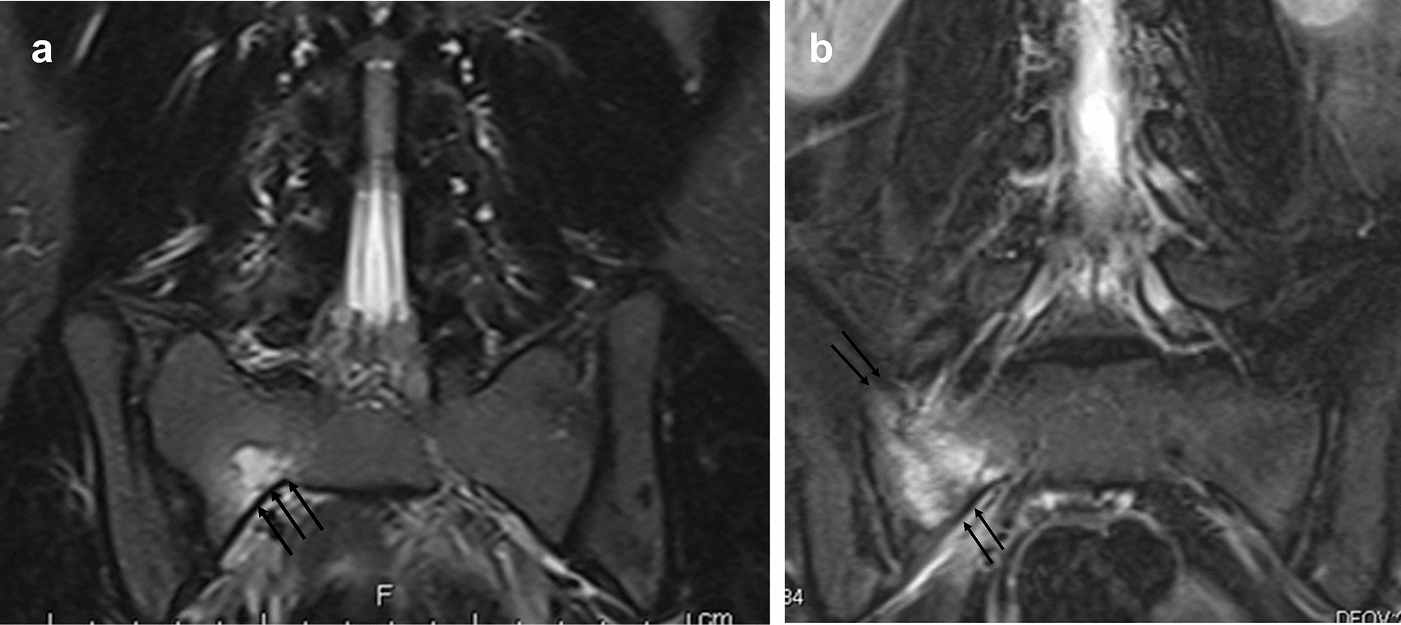
Features Of Sacral Alar Fatigue Fractures In Adolescent Athletes With Overuse Scientific Reports
Diagnostic Accuracy Of Magnetic Resonance Imaging Versus Computed Tomography In Stress Fractures Of The Lumbar Spine Clinical Radiology

Ulnar Stress Fracture Mri Download Scientific Diagram

The Radiology Assistant Stress Fractures

Healthfully Medical Technology Medical Mri Technologist
Stress Fractures In The Foot And Ankle

The Radiology Assistant Stress Fractures

Rit Radiology October 2009 Radiology Medical Imaging Buckle

2 Mri Scan Of Distal Radial Physeal Stress Fracture Note The High Download Scientific Diagram
Posting Komentar untuk "Stress Fracture Mri With Or Without Contrast"