Mri Show Bone Cancer
Each issue is carefully selected to provide a combination of high quality original research informative editorials and state-of-the-art reviews. The MRI is a very sensitive technique that allows better visualization of the bones cartilage and joint tissue.
Giant Bone Island Frontal And Lateral Knee Radiographs Show A Large Well Circumscribed Ovoid Sclerotic Lesion In The Dist Radiology Imaging Radiology Bones

A bone scan can often find bone metastases earlier than an x-ray so it is sometimes used during.

Mri show bone cancer. Click image to align with top of page. Overall 10 of patients with abnormal marrow on MRI were diagnosed with a malignancy. Cancer cells that have spread to the bone can damage the bone and cause symptoms. MRI scans can help medical professionals detect cancer. You might have one to find out whether you have cancer and if you do to measure how big it is and whether it has spread. We provide comprehensive radiology services in Queens New York including bone density tests CT scans dental scans digital mammography digital x.
This test is useful because it. Unlike the current standard screening and detection methods the 3T MRI evaluates the WHOLE of the prostate can ignore the bogus G6 cancer and based upon imaging details in a properly conducted study able to identify reliably with PIRADS 4 and 5 features almost all high-grade cancer anywhere within the prostate. There are several types of bone cancer in dogs. Osteosarcoma arises from bone-forming cells called osteoblasts in osteoid tissue immature bone tissue. This spread is called metastasis. A bone tumor is an abnormal growth of tissue in bone traditionally classified as noncancerous benign or cancerous malignant.
To evaluate damage to cartilage bone or other structures inside and around a joint MRI is the better choice. MRI is also preferred for conditions that. Osteosarcoma is the most common type. This is why treatment may continue even when cancer cells can no longer be seen on an imaging test. Bone marrow is the spongy fatty tissue found inside the bodys larger bones. Tap onoff image to showhide findings.
Health Imaging Services in Westmoreland County. A bone scan is used to check the whole skeleton for bone metastases especially when there is bone pain in several places. Call your Diagnostic Imaging Center in Cooper City Fort Lauderdale Plantation or Coral SpringsMargate today at 954 900-2020. An MRI is a test that produces very clear pictures of the human body without the use of X-rays. This tumor typically occurs in the arm near the shoulder and in the leg near the knee in children adolescents and young adults but can occur in any bone especially. When Cancer Spreads to the Bones.
Bone Density Test. If you find out early that you have a tumor or cancer you can start treatment earlier giving you a head start and improving your prognosis. MRI magnetic resonance imaging is a type of scan that uses magnetism and radio waves to take pictures of inside the body. It has liquid and solid parts. Bone scan A bone scan10 can show if a cancer has spread to other bones and is often part of the workup for people with bone cancer. Bone marrow edema can be seen in a number of different conditions.
An MRI magnetic resonance imaging scan uses magnets to create a detailed picture of your prostate and the surrounding tissues. Cancerous bone tumors usually originate from a cancer in another part of the body such as from lung breast thyroid kidney and prostate. An MRI is a very useful tool for helping your doctors see images of the inside of your body including tissue that cant be seen on a conventional x-ray. Magnetic resonance imaging MRI MRI scans8 create detailed images of the inside of the body using radio waves and strong magnets instead of x-rays so no radiation is involved. Bone cancer and osteosarcoma are not interchangeable terms. An x-ray can also show breaks of the bones.
Instead MRI uses a large magnet radio waves and a computer to. You might also have one to see how well treatment is working. And whole-body magnetic resonance imaging WB-MRI has become available to the general public for cancer screening. The scan takes between 15 and 90 minutes. Get accurate medical imaging from highly experienced board-certified radiologists in NYC with Main Street Radiology. Clinical Oncology is essential reading for all those with an active interest in the treatment of cancerIts multidisciplinary approach allows readers to keep up-to-date with developments in their own as well as related fields.
In many hospitals you may have a special type of MRI scan called a multi-parametric MRI mpMRI scan before having a biopsy. The usual treatment for bone cancer is. T1 weighted image Pathology spine Loss of the normal high signal in the bone marrow indicates loss of normal fatty tissue and increased water content. A bone density test is done with x-rays to check the level of bone minerals in the bone segment. This alone makes up 95 of bone cancer cases. The quantity of the bone mineral content inside bones determines how dense your bones are and the bone density test is a great way to analyze it.
Imaging tests can find large groups of cancer cells but no imaging test can show a single cancer cell or even a few. Let us show you the POM MRI Radiology Centers difference. Incidentally noted abnormal or heterogeneous bone marrow signal on MRI was not. This can help your doctor see if there is any cancer inside your prostate and how quickly. Three patients were later diagnosed with malignancy breast cancer myelodysplastic syndrome merkel cell carcinoma at a median of 19 months. Bone marrow makes these types of blood cellsRed blood cells carry oxygen to all parts of the bodyWhite blood cells help the body fight infection and diseasePlatelets help the blood clot and control bleeding.
There may be a lump pain or neurological signs from pressure. Types of primary bone cancer are defined by which cells in the bone give rise to them. At Excela Health we perform more than 275000 outpatient x-ray procedures a year through a combination of state-of-the-art technologies and attention to patient care and. Racial and ethnic disparities in the use of prostate magnetic resonance imaging following an elevated prostate-specific antigen test. The bone density test usually involves an x-ray of the forearm spine or hip bone. A bone tumor might present with a pathologic.
Abnormal low signal on T1 images frequently indicates a pathological process such as trauma infection or cancer. Before your exam its very important to fill out the safety screening form carefully. In fact it takes millions of cells to make a tumor big enough to show up on an imaging test. An x-ray is usually one of the first tests used to check symptoms like bone pain. Our comprehensive diagnostic imaging services include MRI CT X-Ray Ultrasound and Mammography. It can spread to distant organs such as the lungs.
An MRI picture is in black and white like an XRay but can provide cross sectional pictures from several different perspectives. Published online November 8 2021. Bone cancer develops in the skeletal system and destroys tissue. Cancer that has started in one place can spread to and invade other parts of the body. MRI is safe and painless. If a tumor spreads to the bone its called bone metastasis.
But metal in the scanner can cause serious safety problems or reduce the quality of the images.
Giant Cell Tumour Of Bone Radiology Reference Article Radiopaedia Org Radiology Radiology Imaging Diagnostic Imaging

Pin On Radio Spine

The Radiology Assistant Sclerotic Bone Tumors And Tumor Like Lesions Tumor Bone Diseases Radiology

Pin Op Mri

Pin On Tailbone Mri

Pin On Mri Scans

Pin On Radyoloji

Brain Mri A Systematic Reading Mri Brain Tumor Brain
Pin On Ct Scans

Ct And Pet Scan Vs Mri Pet Scan Mri Ct Scan

Osteoid Osteoma Bony Mass 2 Cm With A Radiolucent Core Osteoid Tumor Radiology Bone Diseases

Pin On Excalibur Healthcare S Imaging Teleradiology Pins

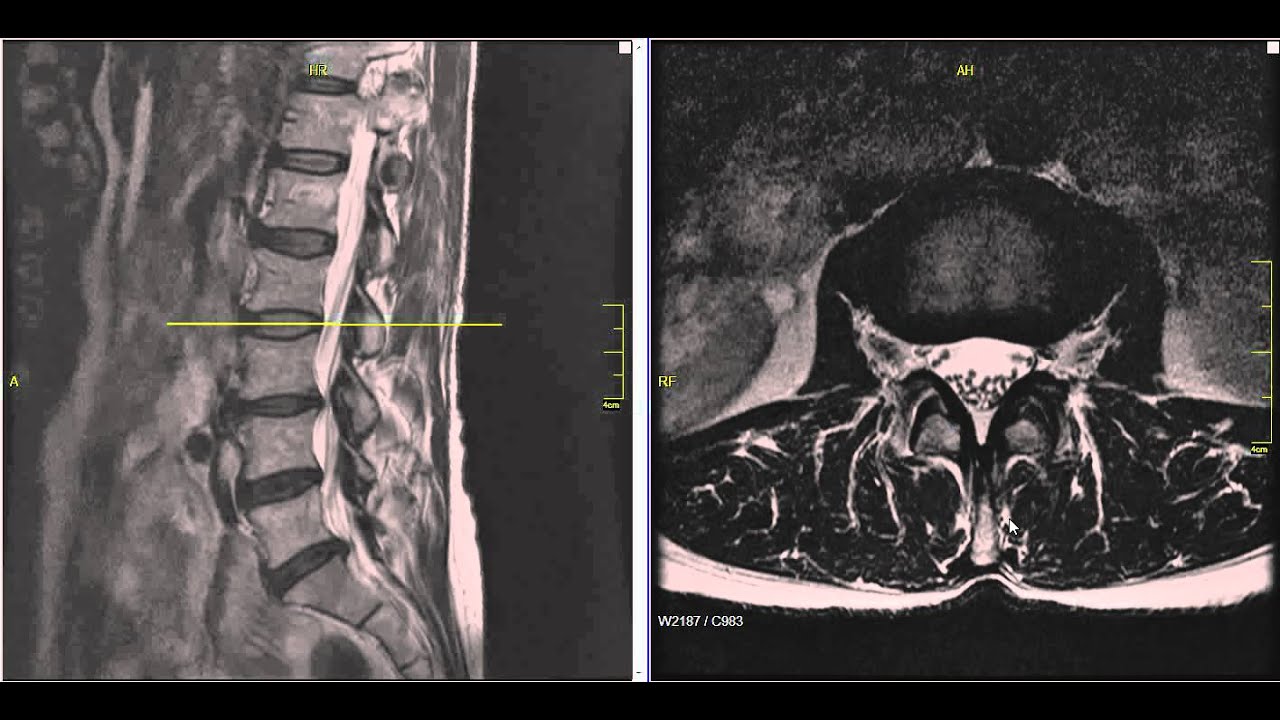
Lumbar Disc Herniation Mri Explained Dr Jeffrey P Johnson Hd Youtube Lumbar Disc Disk Herniation Mri
The Radiology Assistant Sclerotic Tumors In 2021 Tumor Radiology Avascular


Posting Komentar untuk "Mri Show Bone Cancer"