Optic Nerve Meningioma Treatment
Courtesy of Leah Levi MD. Acute loss of vision from an optic neuropathy in a young person is most often due to ON.
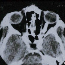
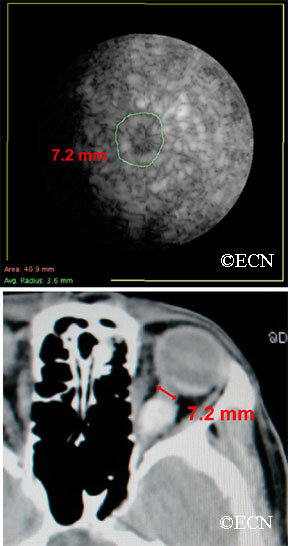
Optic Nerve Sheath Meningioma New York Eye Cancer Center
The optic nerve sheath fenestration is one of the accepted treatment modalities in benign intracranial hypertension.
Optic nerve meningioma treatment. Multiple cranial neuropathies are commonly seen in lesions caused by tumors trauma ischemia and infectionsWhile a diagnosis can usually be made based on clinical features further. Left MRI scan of a sphenoid wing meningioma. Meningioma also known as meningeal tumor is typically a slow-growing tumor that forms from the meninges the membranous layers surrounding the brain and spinal cord. Suprasellar meningioma arises from the base of the skull near the pituitary gland and the optic nerve. In case of malignant gliomas glioblastomas despite treatment including high-dose radiotherapy and chemotherapy these tumors usually result in death within 6-12 months. Specific symptoms due to location.
The tumor has filled the area where the temporal lobe normally lies. Right The illustration shows that as the tumor grows it compresses and displaces normal brain tissue and can encase the arteries and nerves. Symptomatic cavernous angiomas in children usually require treatment due to the high risk of future hemorrhage and greater seizure potential in children. It is a pivotal moment for our world our nation and our brain tumor community. Ask your surgeon about the specific risks of your surgery. If your meningioma cant be completely removed surgically your doctor may recommend radiation therapy following or instead of surgery.
Optic Nerve Sheath Meningiomas ONSM are uncommon benign neoplasms originating from the meningothelial cells of the meninges surrounding the optic nerve. Swelling of the optic disk which is in the retina of the eye where nerve fibers come together to form part of the optic nerve. Located in the spine in some cases against the spinal cord. For instance a meningioma pressing against an optic nerve may cause visual problems. Within the eye there are many elements that work together to create vision. Join David Arons CEO of the National Brain Tumor Society and the entire brain tumor community for an update on the progress we have made in the fight to conquer and cure brain tumors and the critical work that lies ahead in 2021.
Contrast enhancement involving more than half of the length of the optic nerve or continuing into the optic chiasm should arouse the suspicion of neuro-myelitis optica 12. Loss of patches of sight within the field of vision blindness double vision. Optic nerve sheath meningioma and optic nerve glioma may need surgical intervention at times. La diagnosi di meningioma è possibile già alla tomografia computerizzata dove si presenta come una massa a presa di contrasto omogenea e a volte con una o più aree calcificheLa risonanza magnetica con mezzo di contrasto è più utile per definire i rapporti del tumore con le strutture circostanti vasi nervi parenchima cerebrale osso. Cranial nerve palsies can be congenital or acquired. Primary optic nerve meningiomas are less frequent than secondary lesions that extend into the orbit from an intracranial site.
For obese patients weight loss is also needed. Primary ONSM should be differentiated from secondary intracranial meningiomas that. Cranial nerve palsy is characterized by a decreased or complete loss of function of one or more cranial nerves. Electrical disturbances within the brain causing seizures. Outcome The consequences of a hemorrhage from a cavernous angioma are rarely catastrophic in contrast with arteriovenous malformations AVMs or aneurysms. Entrambi gli esami possono evidenziare.
Occasionally seizures dementia trouble talking vision problems one. Treatment for the diplopia associated with abducens nerve palsy can be managed with prisms occlusion botulinum toxin or surgery. Treatment options include medical treatment of the pressure a shunt to drain spinal fluid and lower the pressure andor optic nerve sheath decompression to relieve pressure on the optic nerves. Case 2 Bilateral Vision Loss With Headache. In 1773 John Fothergill was the first to fully describe trigeminal neuralgia in an article presented to the Medical Society of London titled On a Painful Affliction of the FaceIn 1829 Charles Bell distinguished the specific functions of the trigeminal and facial nerves and introduced the idea that the paroxysmal pain in trigeminal neuralgia is directly related to nerve. Pain in the limbs or chest.
The optic nerve is the nerve that connects and transmits information between the eye and the brain. Optic nerve damage is also called optic nerve atrophy or optic neuropathy. Optic nerve damage can lead to vision distortion vision loss and blindness. Symptoms depend on the location and occur as a result of the tumor pressing on nearby tissue. Consider treatment of high-risk patients abnormal MRI with a disease-modifying drug. Symptoms of suprasellar meningioma.
Sixth nerve palsy or abducens nerve palsy is a disorder associated with dysfunction of cranial nerve VI the abducens nerve which is responsible for causing contraction of the lateral rectus muscle to abduct ie turn out the eye. Swelling of the optic nerve head in the back of the eye the first step should be a thorough neurological evaluation followed by radiological studies if needed. The tumor may arise from either the intraorbital or intracanalicular portions of the optic nerve where there is a meningeal sheath. They are the second most common optic nerve tumor accounting for 2 of space-occupying orbital masses. The treatment of optic nerve gliomas is controversial. The inability of an eye to turn outward results in a convergent strabismus or esotropia of which the primary symptom is diplopia commonly.
Visual prognosis is excellent with ON. Table 4 Treatment choices after surgery by extent or no excision if surgery was not possible for different kinds of meningioma. An optic nerve sheath meningioma can look exactly like optic neuritis on MRI and should be suspected if the contrast enhancement does not subside within 3 months. Optic nerve sheath meningiomas derive from the arachnoid sheath of the optic nerve. Symptoms of spinal meningioma. No treatment may be required in patients with no growth good vision no cosmetic deformity.
The Canadian Journal of Ophthalmology CJO is the official journal of the Canadian Ophthalmological Society and is committed to timely publication of original peer-reviewed ophthalmology and vision science articles. Occlusion using a Bangerter filter or patch can eliminate diplopia and confusion prevent amblyopia or suppression in children and decrease the possibility of ipsilateral medial rectus contracture. Patients for whom treatment carries a. Optic Nerve Sheath Meningioma. Many cases never produce symptoms. When a meningioma does recur it may be the same grade or a more aggressive or malignant form.
Completely excised Simpson 1 to 3 Incompletely excised Simpson 4 to 5 No excision radiological only diagnosis Recurrent. The assessment of MS risk is based on the brain MRI. In general there are three basic options. Any meningioma may come back. For instance surgery to remove a meningioma that occurs around the optic nerve can lead to vision loss. Tumors in this area can cause visual problems and dysfunction of the pituitary gland.
Numbness and weakness or the arms. The decision of whether to and how best to treat a meningioma is based on multiple factors including size and location of the tumor symptoms growth rate and age of the patient among others. Another tumors location may affect motor skills or speech.
Pin On Meningioma Edu For You

Diagnosis And Management Of Optic Nerve Sheath Meningiomas Sciencedirect

Optic Nerve Sheath Meningioma Statpearls Ncbi Bookshelf
Pin On Genealogy Clips

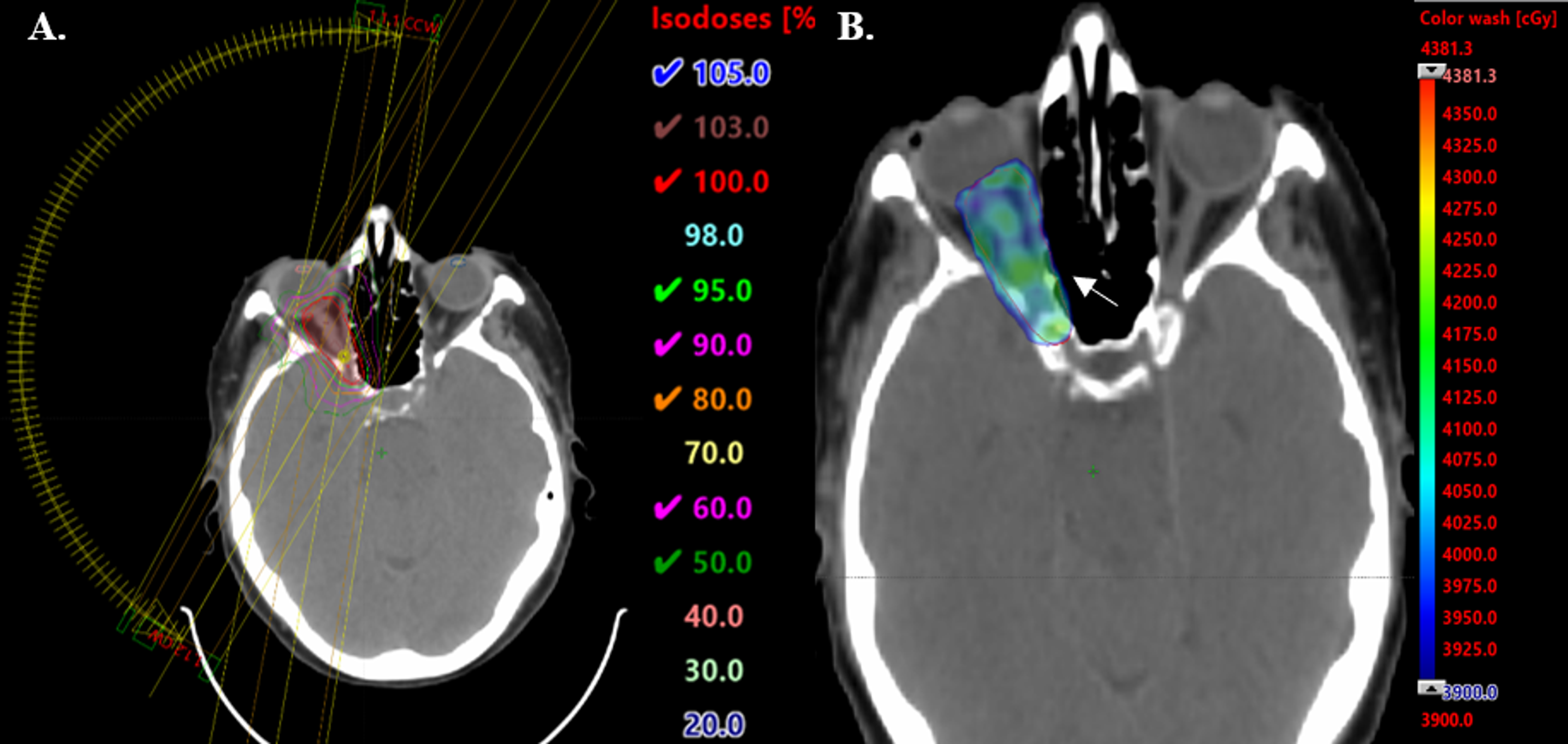
Cureus Salvage Radiosurgery For Optic Nerve Sheath Meningioma

Pin On Brain

Diagnosis And Management Of Optic Nerve Sheath Meningiomas Sciencedirect

Cureus Treatment Of Recurrent Optic Nerve Sheath Meningioma With A Secondary Course Of Radiotherapy
Pin On Ct

Optic Nerve Sheath Meningiomas Non Surgical Treatment Clinical Oncology

Cureus Treatment Of Recurrent Optic Nerve Sheath Meningioma With A Secondary Course Of Radiotherapy
Optic Nerve Sheath Meningioma New York Eye Cancer Center
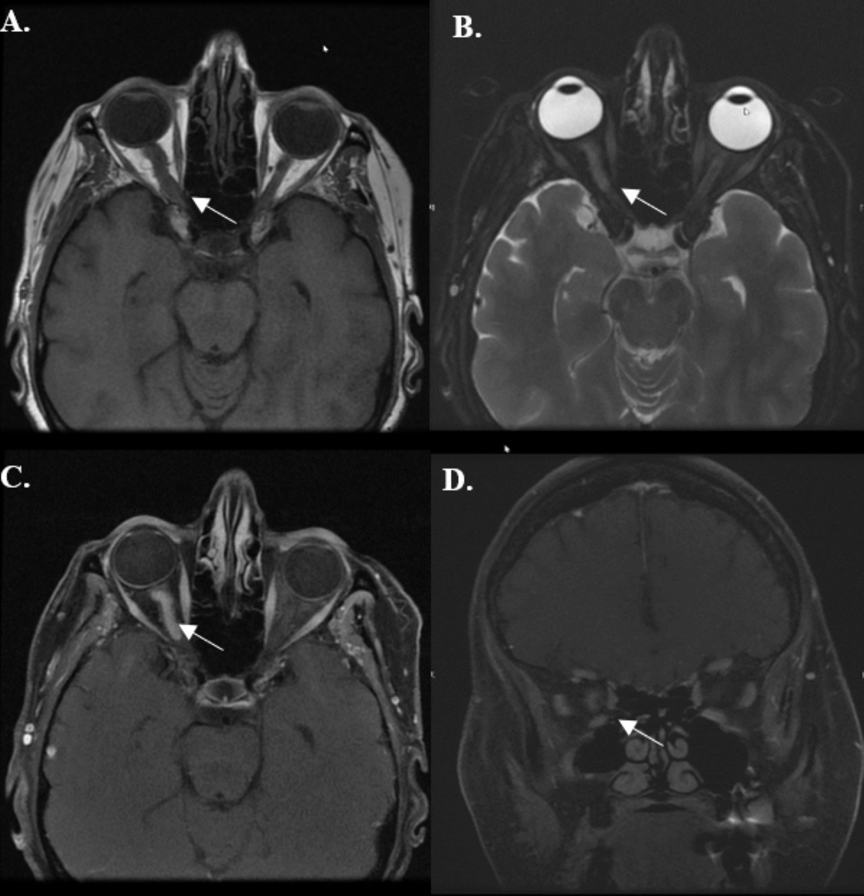
Intracanalicular Optic Nerve Meningioma A Serious Diagnostic Pitfall American Journal Of Neuroradiology

Intracanalicular Optic Nerve Meningioma A Serious Diagnostic Pitfall American Journal Of Neuroradiology


Posting Komentar untuk "Optic Nerve Meningioma Treatment"