Diagnostic Nerve Root Block
The sciatic nerve is the longest nerve in the human body with nerve root L4 L5 S1 S2 S3 and is the continuation of. Electromyography or EMG is used to diagnose nerve and muscle disorders spinal nerve root compression and motor neuron disorders such as amyotrophic lateral sclerosis.
Southwest Health Sensory Nerve Root Block Snrb
A systematic review in the field of clinical diagnostic of disc herniation with lumbar nerve root involvement NRI has terminated the search of literature at October 2008 and an update is in progress.
Diagnostic nerve root block. A total of 75 ON patients who underwent CDR from 1998 to 2012 were included in this analysis. Diagnostic Occipital Nerve Blocks. Sensory nerve biopsy is an established diagnostic procedure. Coding Billing for Medial and Lateral Nerve Blocks. Muscles develop abnormal electrical signals when there is nerve or muscle damage. This is usually because of the lingering effects of the nerve ablation.
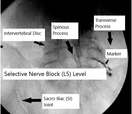
N Suite 100 Maple Grove MN 55369 for all of your CT Injections Biopsies MRI X-ray and other diagnostic imaging needs. Visit RAYUS Radiology Maple Grove MN at 9630 Grove Cir. 55 patients were included because they met the International Headache Societys IHS diagnostic criteria for ON responded to CT-guided nerve blocks at the C2 dorsal nerve root and had at least 1 follow-up visit. After the procedure you may still feel pain for up to 14 days. In 5 studies mean pain reduction ranged from 82 at 2 weeks after the first block to -01 at 1 month after the third block. A patient was seen at our facility and underwent a left-sided L5 and S1 S2 S3 and S4 lateral branch nerve block for diagnostic purpose with C-arm fluoroscopy.
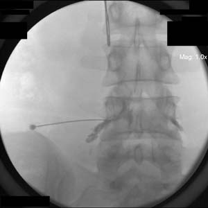
This website features single shot and continuous nerve block techniques. Therefore no search of the literature was performed by the present authors. What are the correct codes for a lateral nerve block. Fluoroscopic guidance helps improve the diagnostic accuracy and decreases procedural risks. Total Hip Replacement - nerve compression and stretch during surgery causing damage to the sciatic nerve that serves the majority of muscle groups in the lower limb resulting in dysfunction. Nerve conduction studies NCS.
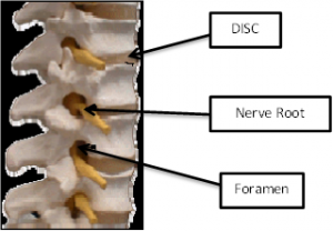
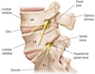
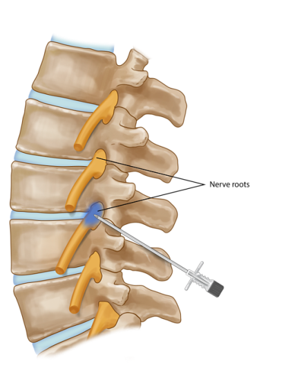
Injections may also be used diagnostically as a selective nerve block to confirm the nerve root as the cause of the leg pain and is typically helpful prior to. Grade 3 scars correspond to symptoms and pain more than do Grades 1 and 2. Three studies reported minor. A systematic review in the field of clinical diagnostic of disc herniation with lumbar nerve root involvement NRI has terminated the search of literature at October 2008 and an update is in progress. Regional Anesthesia Ultrasound has become a recent standard to perform regional anesthesia. A nerve block might also be used as a diagnostic tool to find out where your pain is originating from.
Therefore no search of the literature was performed by the present authors. Atypical trigeminal neuralgia ATN is a rare form of neuralgia and may also be the most misdiagnosed form. Difficulties arise when multiple mononeuropathies become confluent thus making the differentiation from polyneuropathy difficult. Pediatric Neurology publishes timely peer-reviewed clinical and research articles covering all aspects of the developing nervous systemPediatric Neurology features up-to-the-minute publication of the latest advances in the diagnosis management and treatment of pediatric neurologic disorders. The Medial Branch Block MBB and Radiofrequency Ablation RFA are 2 separate procedures used to diagnose and treat your pain. Delayed conduction prolonged distal latency conduction block andor slow conduction velocity across the lesion but normal conduction distal to the lesion.
The procedure is primarily diagnostic meaning that if the patient has the appropriate duration of pain relief after the medial branch nerve block then he or she may be a candidate for a subsequent procedure - called a medial branch radiofrequency neurotomy or ablation - for longer term pain relief. If three 3 medial branch nerves are injected only two 2 facet. By seeing how a nerve block affects your pain your doctor may be able to determine the. Disc herniation with nerve root involvement. Normal spontaneous activity but may show decreased motor unit action potential MUAP recruitment due to conduction block. The symptoms can be mistaken for migraines dental problems such as temporomandibular joint disorder musculoskeletal issues and hypochondriasisATN can have a wide range of symptoms and the pain can fluctuate in intensity from mild aching to a crushing.
This procedure can relieve symptoms associated with dorsal scapular nerve entrapment on a long-term basis. Disc herniation with nerve root involvement. A medial branch block is similar but the medication is placed outside the joint space near the nerve that supplies the joint called the medial branch a steroid may or may not be used. The journals editor Yasmin Khakoo MD FAAN in conjunction. A Grade 3 scar may also extend to the nerve root whereas Grades 1 and 2 do not. Dorsal scapular nerve ablation Pulsed radiofrequency treatment Pulsed radiofrequency lesioning of the dorsal scapular nerve is indicated in patients who show a positive response to the diagnostic dorsal scapular nerve block.
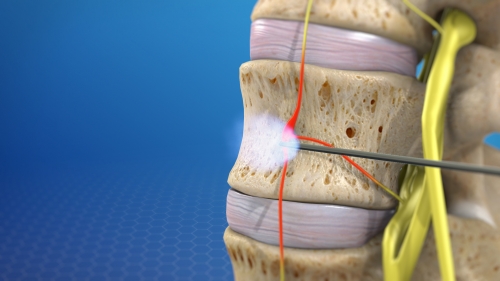
The DRG stimulator is well-suited for specific neuropathic conditions which include CRPS pelvic pain groin neuralgia extremity pain and other nerve pain involving isolated parts of the body. G548 Other nerve root and plexus disorders - intercostal neuritis Infraclavicular-Suprascapular Nerve Blocks 64415 Injection. EDx features of nerve conduction slowing or block. We begin with the MBB which is used as a diagnostic tool to establish if the source of your pain are the facet joints and the medial branch nerves. Radiofrequency Nerve Ablation is Proven to Be Effective for Nerve Pain. Radiofrequency nerve ablation is an option available to those who have received a diagnostic pain receptor block injection.
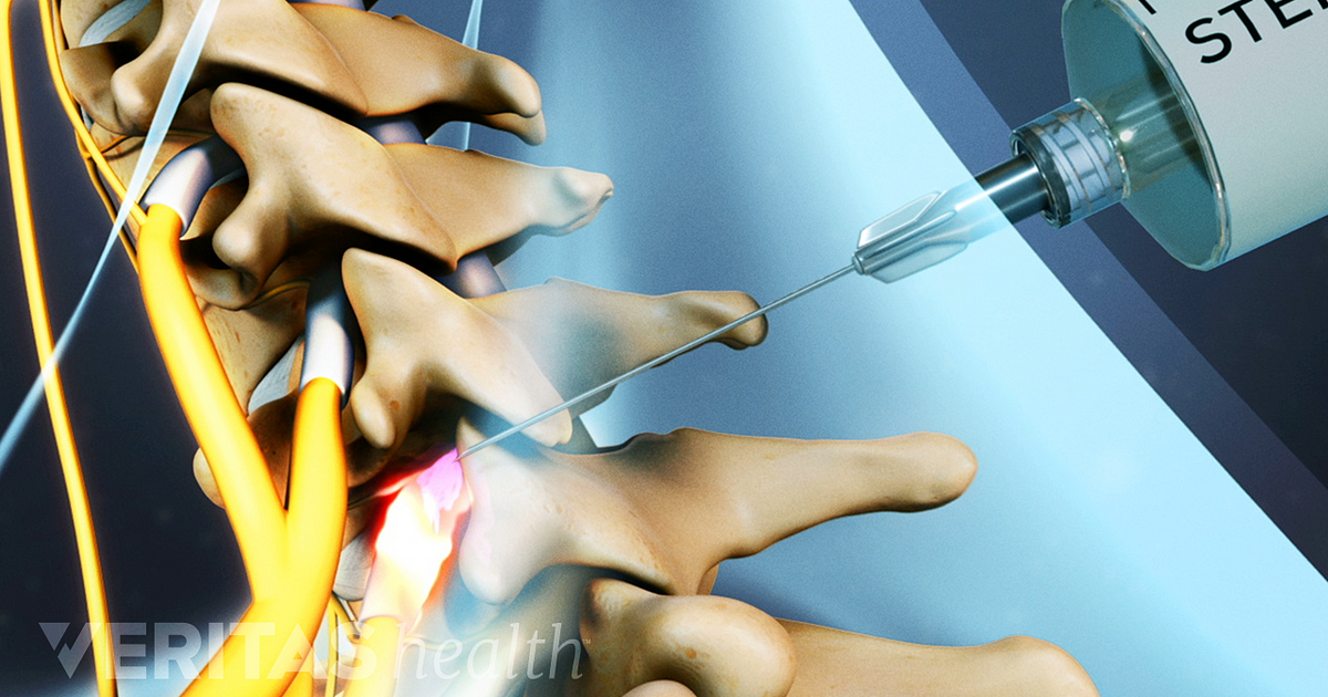
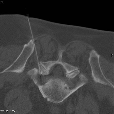
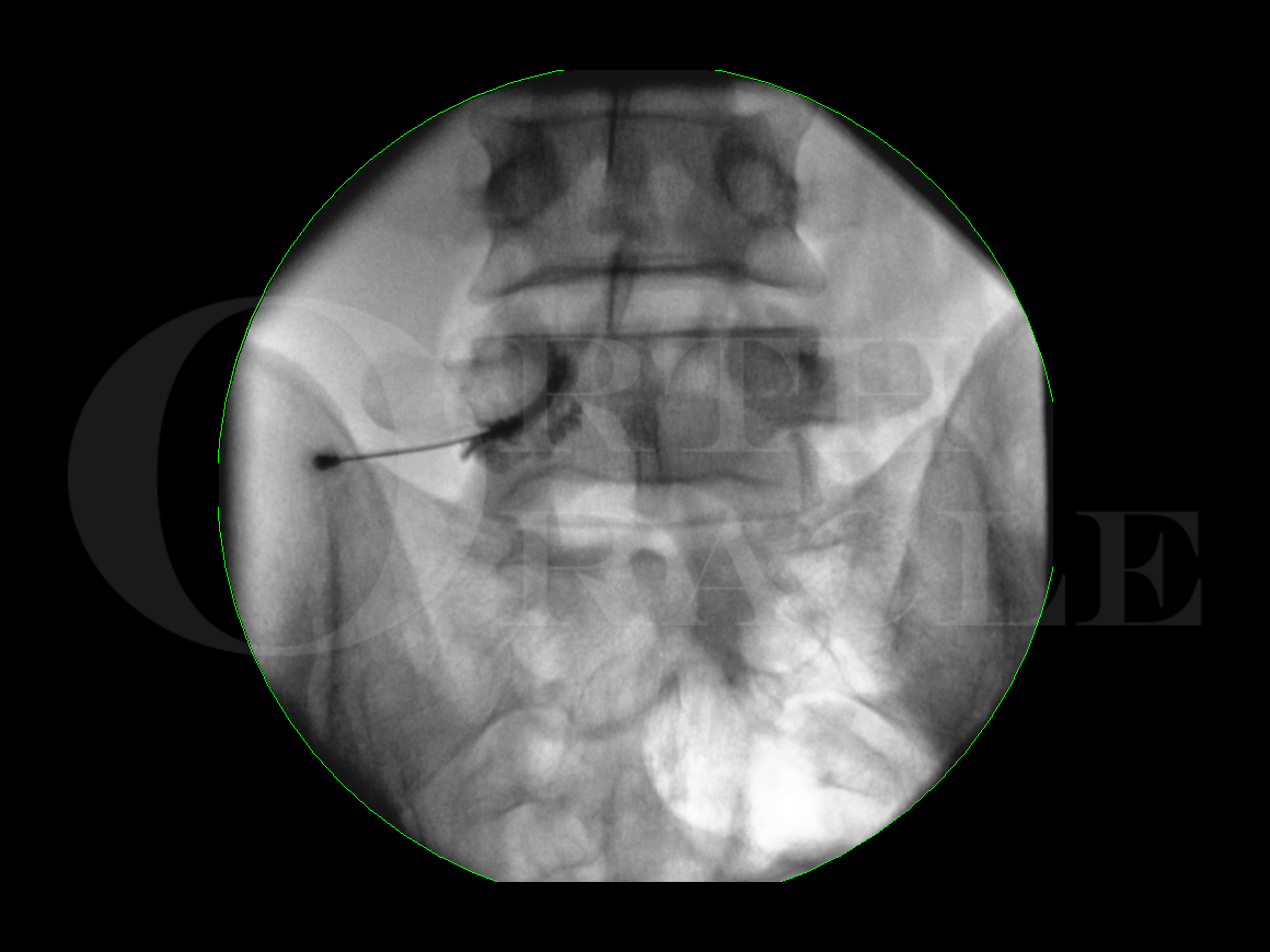
When Dorsal Root Ganglion therapy should be considered. Grade 1 scars tend to be mild and made up of thin fibrous bands that are laid down over the dura mater which is the outermost spinal cord covering described above. Clinically Relevant Anatomy edit edit source. A fluoroscope or low-powered X-ray allows whoever administers the nerve block to visualize the bony structures. Reported at a level of 1. A facet block is an injection of local anesthetic and steroid into a joint in the spine.
Facet joints are small bony projections from one vertebra which meet. According to the AMA the code series for medial branch blocks and the facet joint injections are the same ie CPT series 64490-64495 with reporting based on the number of facet joints injected not the number of nerves injected. Surgical decompression of second cervical nerve root and ganglion Surgical decompression of the greater occipital nerve. EMG records the electrical activity in the muscles. Thoracic paravertebral block TPVB is the technique of injecting local anesthetic alongside the thoracic vertebra close to where the spinal nerves emerge from the intervertebral foramen. Muscle power testing in the context of nerve and root distribution is crucial.
And 23 after 35 months. This produces unilateral segmental somatic and sympathetic nerve blockade which is effective for anesthesia and in treating acute and chronic pain of unilateral origin from the chest and.
Selective Nerve Blocks Sapna Spine And Pain Clinic Of North America Fairfax Va
About Nerve Blocks Pain Management Henry Ford Health System Detroit Mi

Selective Nerve Root Block Complete Orthopedics Multiple Ny Locations
Selective Nerve Root Block Legacy Spine Neurological Specialists
Nerve Root Block Ct Guided L5 Radiology Case Radiopaedia Org
Selective Nerve Root Block Perineural Injection

Understanding Diagnostic Injections Selective Nerve Root Block Snrb
Selective Nerve Root Block Legacy Spine Neurological Specialists
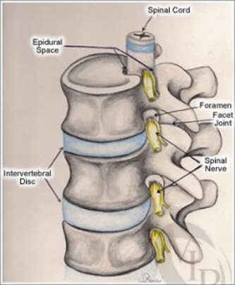
Fig 3 Selective Cervical Nerve Root Blockade Experience With A Safe And Reliable Technique Using An Anterolateral Approach For Needle Placement American Journal Of Neuroradiology

Selective Nerve Root Block Injections
Lumbar Nerve Root Block Surgical Technique Orthoracle
Selective Nerve Root Block Indications And Applications Bone And Spine
Nerve Root Blocks Imaging Glossary Patients Ur Medicine Imaging Sciences Radiology University Of Rochester Medical Center
Selective Nerve Root Block Plano Tx Texas Back Institute


Posting Komentar untuk "Diagnostic Nerve Root Block"