Lumbar Spine X Ray Positioning Standing
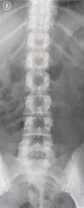
The science of biomechanics helps explain the causes of cell tissue organ and body system disorders and supports clinicians in the diagnosis prognosis and evaluation of treatment methods and. The conus is in normal position at the thoracolumbar junction.
Functional Anatomy And Radiology Of The Spinal Column Clinical Gate
Dual-energy radiographic x-ray absorptiometry available since 1987 is the current standard of care for bone mass measurement.

Lumbar spine x ray positioning standing. Lumbar Spine AP or PA. If they need to do further tests to examine the cause of the lumbar strain the doctor may request. My back pain seems to be only on my left side by my ribs. On a 15 Tesla magnet multiple sagittal and axial images were performed through the lumbar spine using variable pulse sequences. The effects of staff positioning different X-ray imaging systems and freestanding radiation protection shields RPSs were considered. Slight narrowing of the disc space between C5-6.
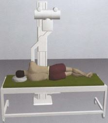
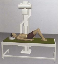
In trauma patients a lumbar spine X-ray is done in the AP or PA position with minimal movement of. In a type III dens fracture the fracture line extends into the body of the C2 vertebra. It uses an x-ray tube instead of an isotope to generate dual energy photons resulting in higher image resolution and greater speed than DPA. Even this can be problematic though. Preparing for lumbar spinal fusion Overview. A lateral cervical spine x-ray was subsequently obtained after the patient was placed in a halo traction device.
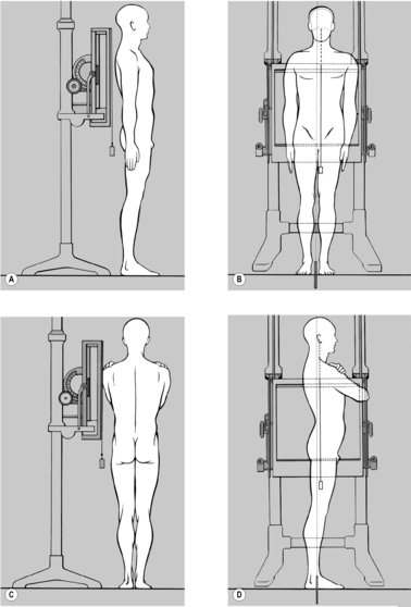
Move slider bar so as to snugly rest under right arm. Journal of Manipulative and. Abnormal movement of the vertebrae rubbing against one another may result in back leg or arm pain. The Scoliosis X-ray is the most common method of diagnosing and monitoring a patients scoliosis. Remove any artifacts in the desired field eg clothing with hooks snaps zippers. Radiography is an imaging technique using X-rays gamma rays or similar ionizing radiation and non-ionizing radiation to view the internal form of an objectApplications of radiography include medical radiography diagnostic and therapeutic and industrial radiographySimilar techniques are used in airport security where body scanners generally use backscatter X-ray.
He noted a more reliable correlation between anatomic and X-ray measurements of pelvic incidence than of sagittal. A doctor is likely to perform a physical examination to diagnose a lumbar strain. Standing behind the patient place base bar of calipers under left arm. My x ray says that i have a right curvature of the thoracic spine from the superior T6 vertebral body to T11 vertebral body that measures 15 degrees Also a Left curvature of the lumbar spine from superior T12 to inferior L4 measures 17 degrees. Coccydynia occurs in the lowest part of the spine the coccyx which is believed to be a vestigial tail or in other words the tail boneThe name coccyx is derived from the Greek word for cuckoo due to its beak like appearance. An X-ray can help determine the amount of curvature in the spine.
Position of patient Supine or prone. In the standing position the mean pelvic tilt angle which is open at the back is 13 6. The content on or accessible through Physiopedia is for informational purposes only. Objectives To reduce occupational radiation exposure in a hybrid operating room OR used for three-dimensional 3D image guided spine procedures. Injured patients should NOT be turned over. Methods An anthropomorphic phantom was imaged with a robotic ceiling.
A thorough medical examination including X-ray and in some cases CT or MRI scans are necessary to diagnose bulging disc conditions to the full extent accurately. 1 is one of the oldest physiotherapy schools in France and one of the first schools to be integrated into the Faculty of Medicine program in LyonPhysiotherapy is an integral part of. The Spine Journal is the 1 ranked spine journal in the Orthopaedics category. Functions of the low back or lumbar area include structural support movement and protection of certain body tissues. No suspect bone marrow lesions are present. Pediatric Spondylolysis Spondylolisthesis represent a continuum of disease where there is a fracture of the pars interarticularis spondylolysis which may progress to anterior subluxation of one vertebral body anterior to the adjacent inferior vertebral body spondylolisthesis.
Activities that place stress and strain on the spine can lead to the weakening of the discs. Then I had another appointment with him about my neck hes sending me to get an MRI done but so far on X-ray is. Once any underlying pathology or conditions are ruled out X-rays can tell us what we need to know about a patients conditionWhile many people still have concerns regarding side effects of frequent X-rays those concerns are unfounded. The Lyon school of physiotherapy for scoliosis managed by Dr. There is normal lumbar alignment. A 2014 study published in Spine Journal found that positioning during testing and the number of vertebrae included in the measurement may contribute to skewed results.
Jean Claude de Mauroy the head of the orthopedic medicine department at Clinique du Parc Lyon France Fig. Purpose and Structures Shown A basic view of the lumbar spine. I have an S curved spine. The spine may straighten while lying down in people with postural kyphosis. These testing formats show changes in shape and condition of the disc. Place patient in gown.
Lumbar fusion physical therapy post op protocol n psifaliflliftlif typical psif can be done alone or in combination with anterior lateral lumbar or transforaminal lumbar interbody fusion placement of one or multiple interbody devices into disc space through posterior anterior lateral or transforaminal approach. Spinal fusion is a surgery that permanently joins together one or more bony vertebrae of the spine. An X-ray is the first step healthcare providers take to measure the lordotic curve. If your doctor wants more detailed images they may. Validity and Reliability of Standing Posture Measurements Using a Mobile Application Hopkins et al. Clinical Biomechanics is an international multidisciplinary journal of biomechanics with a focus on medical and clinical applications of new knowledge in the field.
The Spine Journal the official journal of the North American Spine Society is an international and multidisciplinary journal that publishes original peer-reviewed articles on research and treatment related to the spine and spine care including basic science and clinical investigations. MRI OF THE LUMBAR SPINE History. Coccydynia is also known as coccygodynia coccygeal pain coccyx pain or coccalgia. A Lateral cervical spine x-ray. This patient was noted to have a type III dens fracture on CT scan. The position of the lumbar spine attached to the sacral plateau is thus affected by the pelvic tilt and by the sacral slope.
Pain in the low back can be a result of conditions affecting the bony lumbar spine intervertebral discs discs between the vertebrae ligaments around the spine and discs spinal cord and nerves muscles of the low back internal organs of the pelvis. Cervical spine x-ray today shows there is flattening of the cervical lordosis. Physiopedia is not a substitute for professional advice or expert medical services from a. Secure lead apron around patient. Lateral Thoracic Spine.
Ce4rt Radiographic Positioning Of The Lumbar Spine For X Ray Techs
Position Of Taking Hanging Total Spine X Ray Hanging Total Spine X Ray Download Scientific Diagram

L S Spine Ap Lat Anatomy And Physiology Part 13 Youtube

Lumbar Spine Radiographic Anatomy Diagnostic Imaging Radiology Imaging Radiology Student

Position Of Hanging Total Spine X Ray Hanging Total Spine X Ray Was Download Scientific Diagram
Thoracic Spine Radiographic Anatomy Radiology Radiology Student Radiology Imaging

Ce4rt Radiographic Positioning Of The Lumbar Spine For X Ray Techs
The Lowdown On Lumbar Spine Positioning

Pin On X Ray Vision

Xray Of Whole Spine Ap Lateral Position In Dark Background Stock Photo Download Image Now Istock
Pin By Kareena Summers On Medical Imaging Radiology Student Radiologic Technology Radiography

Why Should Lumbar Spine Radiographs Be Obtained With The Patient In The Standing Position Whenever Possible Spine Secrets

Ce4rt Radiographic Positioning Of The Lumbar Spine For X Ray Techs
Boning Up On Humerus Clavicle And Ac Joint Positioning Diagnostic Imaging Radiology Radiology Student


Posting Komentar untuk "Lumbar Spine X Ray Positioning Standing"