Acute Infarction Brain Mri
The International Journal of Cardiology is devoted to cardiology in the broadest senseBoth basic research and clinical papers can be submitted. MRI does not involve X-rays or the use of ionizing radiation which distinguishes it from.

Pin On Understand Your Body
Cerebral edema can occur with acute hypoxic encephalopathy large cerebral infarction and severe traumatic brain injury.
Acute infarction brain mri. A stroke can also occur if a blood vessel inside the brain bursts leading to bleeding inside the head. International Journal of Cardiology is a transformative journal. Symptoms may include headache lethargy fever and focal neurologic deficits. On T2-weighted and FLAIR images ischemic infarction appears as a hyperintense lesion usually seen within the first 38 hours after stroke onset Figure 1 2325In a recent study of acute ischemic stroke patients studied by MRI within 6 hours of symptom onset patients without a visible hyperintense lesion on FLAIR. Restricted diffusion in a wedge-shaped region of the brain arrow is a characteristic finding of a recent cerebral infarct. There are two main types of stroke.
Read more if the infection penetrates into brain parenchyma. A stroke is a medical condition in which poor blood flow to the brain causes cell death. Imaging plays a key role in determining the most probable diagnosis pointing to the next steps of investigation and providing prognostic. Cerebral herniation occurs when the brain is displaced under the cerebral falx the cerebellar tentorium or through the foramen magnum causing loss of brain stem functions fig. Hesitating speech transcortical aphasia. Brain cells can die causing lasting damage.
T2 and Fluid Attenuated Inversion Recovery FLAIR imaging. Midline shift towards left in a right-sided subdural hematoma chronic subdural hematoma with an acute bleeding component. Toxic and metabolic brain disorders are relatively uncommon diseases that affect the central nervous system but they are important to recognize as they can lead to catastrophic outcomes if not rapidly and properly managed. Axial sagittal and coronal see the example image below. Magnetic resonance imaging MRI is one of the most commonly used tests in neurology and neurosurgeryMRI provides exquisite detail of brain spinal cord and vascular anatomy and has the advantage of being able to visualize anatomy in all three planes. Multiple embolic infarction diffusion and FLAIR imaging.
A stroke is sometimes called a brain attack If blood flow is cut off for longer than a few seconds the brain cannot get nutrients and oxygen. MRI scanners use strong magnetic fields magnetic field gradients and radio waves to generate images of the organs in the body. Diagnosis is by contrast-enhanced MRI or CT. Risk factors for malignant MCA syndrome. Cerebrovascular Disease stroke or brain attack. Signs and symptoms of a stroke may include an inability to move or feel on one side of the body problems understanding or.
Ischemic due to lack of blood flow and hemorrhagic due to bleeding. However in the acute phase mortality rate is approximately 10 to 20. Magnetic resonance imaging MRI is a medical imaging technique used in radiology to form pictures of the anatomy and the physiological processes of the body. However it may be adjusted by mass lesions or in the setting of cerebral edema. The physiologic volume of the brain parenchyma is a relatively constant value in adults. Brain abscess Brain Abscess A brain abscess is an intracerebral collection of pus.
In contrast to MS ADEM favors subcortical and deep white matter regions without Dawsons fingers or involvement of the callosal-septal interface. Learn more about APCs and our commitment to OA. It is important to note that mixed-attenuation SDHs are not necessarily acute on chronic. Areas of high signal on the DWI images and low signal on the ADC images indicate restricted diffusion - an indicator of a pathological process of cell death such as infarction cancer or abscess formation. 28 On MRI multiple bilateral and usually asymmetric T2-hyperintense lesions will be present Figure 15. Proximal MCA infarction or cerebellar infarction may lead to marked edema causing herniation and death eg malignant MCA syndrome.
Both cause parts of the brain to stop functioning properly. Speaks nonsense words fluent aphasia time-lapse movies Acute stroke. Edema usually peaks after 3-4 days but reperfusion may accelerate this. Shutter 2019 Decompressive craniectomy may prevent this by opening of a generous bone window. And infarction MRI has substantially higher sensitivity than CT for the detec-tion of EDH SDH nonhemorrhagic cortical contusions brain - stem injuries and white matter axonal injuries supported by level Ib evidence. The journal serves the interest of both practicing clinicians and researchers.
Magnetic Resonance Imaging MRI of the Brain and Spine. Writes but cant read alexia without agraphia Subacute stroke.

White Matter Diseases White Matter Radiology Imaging Radiata

Acute Infarction Dwi Adc Occipital Lobe Intense Image Shows

Fd Acute Left Mca Territory Infarction Mca

The Radiology Assistant Brain Ischemia Imaging In Acute Stroke Mri Brain Mri Radiology

Hyperacute Infarction 6 Hrs Hyperdense Mca Sign Mca Signs False Positive

Pin By Andres Sanchez On Radiology Brain Images Nervous System Anatomy Radiology Imaging

55 Yr Male Left Sided Weakness Dx Acute Ischemic Infarction In The Right Parietotemporal Lobes In The Territory Of Mca
Stroke Evolution From Acute To Chronic Infarction Radiology Video Tut Radiology Mri Mri Brain

Bilateral Pca Territory Infarction Different Ages Radiology Case Radiopaedia Org Radiology Radiology Imaging Brain Anatomy

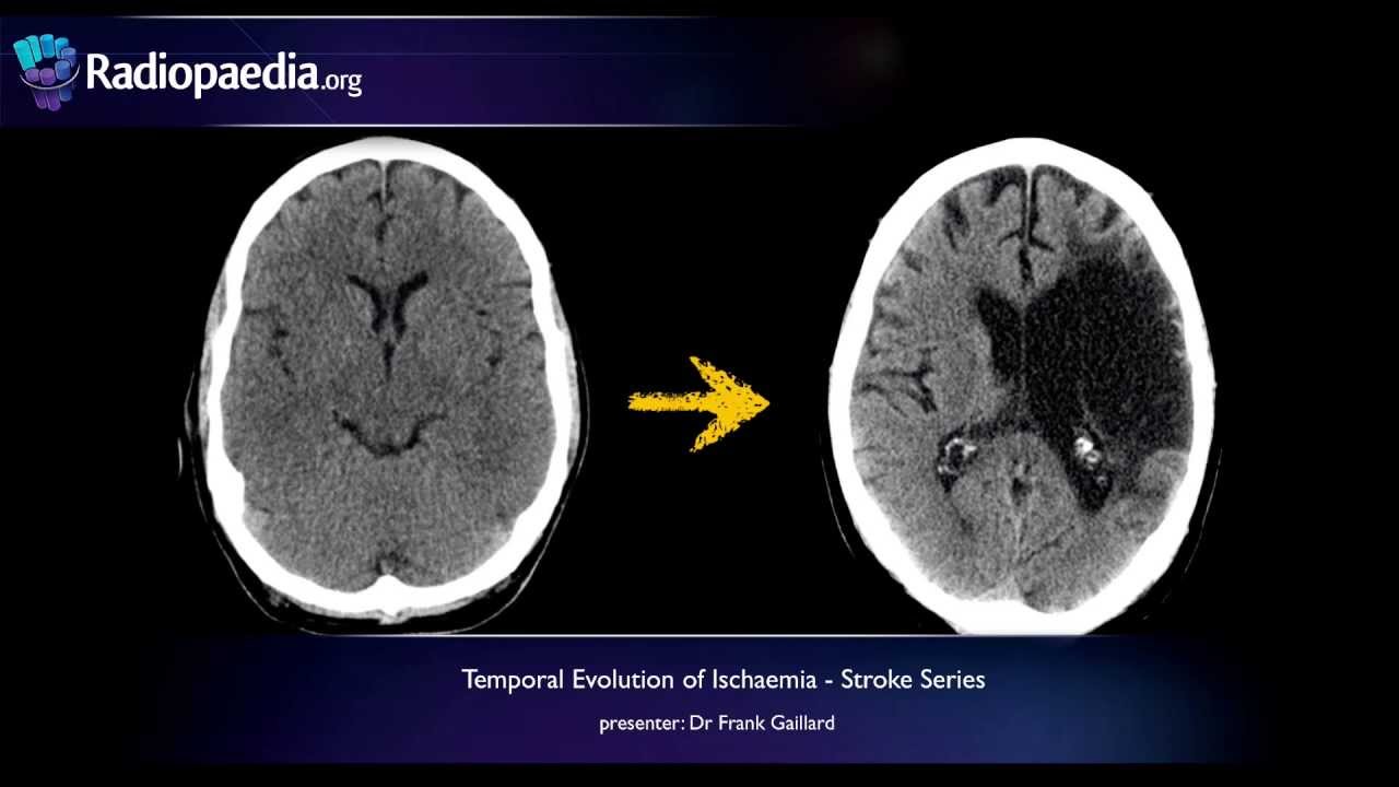
Stroke Evolution From Acute To Chronic Infarction Radiology Video Tutorial Ct Mri Youtube Brain Scan Ct Scan Brain System

Imaging Acute Stroke Youtube Radiology Imaging Radiology Medical Laboratory Science

Stroke Evolution From Acute To Chronic Infarction Radiology Video Tutorial Ct Mri Youtube Radiology Pet Ct Sonography

Acute Infarction Along The Left Anterior Choroidal Artery Distribution Abnormal Signal And Diffusion Carotid Artery Internal Carotid Artery Internal Capsule

Pin On Ct Head
Posting Komentar untuk "Acute Infarction Brain Mri"