Pituitary Mri What To Expect
MRI uses a magnetic field and radio waves to create detailed images of the organs and tissues within your body without the use of ionizing radiation. It is well lit and there is a fan for patient comfort.
How To Read An Mri Of The Pituitary Gland Youtube

MRI technology uses a magnetic field radio waves and a computer to create detailed image slices cross sections of the head.
Pituitary mri what to expect. So the next step my endocrinologist is taking is IPSS with the Interventional Radiology department at the Army hospital here. The pituitary stalk is slightly deviated towards the left. An MRI offers a much clearer picture and involves being in a more confined space than a CT scanner. Depending on what your doctor is looking for this test may be ordered with or without IV contrast. The pituitary is a very small hormone-producing gland located behind the nose. Lie Down on the MRI Table.
A pituitary MRI or magnetic resonance imaging is a technique that takes pictures of the brains pituitary gland and its surrounding areas. I was prescribed bromocriptine. First medical technologists will instruct you to remove and store any metal materials in a lockable storage area. Thyroid hormone and adrenal steroids can be taken as pills. There is a CSF intensity region involving the upper posterior portion of the pituitary gland measuring 5x2 mm which does not enhance. Dec 13 2013 at 846 PM.
Learn how to detect and diagnose the serious symptoms of these pituitary tumors right now. There is also a two way intercom system for communication between patient and technologist. Magnetic Resonance Imaging MRI is a safe as well as painless procedure. Pediatrician told me that he might possibly have a pituitary or pineal tumor that could be large enough to affect his vision. This is the electric current in the scanner coils being turned on and off. If the ACTH is very low an adrenal tumor or use of steroid-containing medications is more likely.
MRI images are usually more detailed than those from CT scans see below. My pituitary MRI did not show a adenoma. After the MRI you go into a special treatment room. Image_slider The pituitary gland is a small gland that controls the bodys hormones1. If the workup indicates a pituitary tumor or disease many clinicians seek the help of pituitary disease experts. The first thing to consider is whether the patients symptoms that led to the MRI are related to the tumor.
The part of the body being scanned will be placed in the middle of the magnet. Hey girls so I finally convinced my husband to do a semen analysis and results were normal. Hi I had an MRI that says this and I am waiting to see the endo but am wondering if I could get any insight into what to expect. MRI scans will be the main follow-up tests along with testing hormone levels if your tumor made hormones. Here are the results. Having a scan is painless and will not harm you.
If the presenting symptoms are related to mass effect from the pituitary tumor apoplexy visual loss cranial nerve dysfunction or hormone excess or deficiency then the lesion is not an incidentaloma and. Using the points mapped from the MRI several narrow beams of high-dose radiation are delivered to. These people will need hormone replacement. The scan locates the precise location and size of the tumor. Youre usually able to listen to music through headphones during the scan if you want to and in some cases you can bring your own CD. After doing some research I found this forum and got some really good advice.
Next cycle I will be doing IUI with clomid. If the biochemical testing points to a pituitary source. But my prolactin was elevated and MRI show a pituitary tumor 5mm. Immediately after transnasal transsphenoidal pituitary gland surgery you will recover in an intensive-care unit of the hospital where your healthcare team will monitor your heart rate blood pressure and respiration as you recover from general anesthesia. Enter the Scanning Room. The MRI scanner will make loud tapping noises at certain times during the procedure.
I have a consult with that department on 05172016 and will be scheduled for the actual procedure later that week or the next. Then came the dizzy spells and morning sickness. Positive imaging findings were observed in 84 3137 of the subjects. In the modern era of advanced imaging they are a common finding. Benign pituitary tumor. What to Expect With a Brain MRI.
The endocrinologist may want you to have a scan of the pituitary gland using an MRI or CT scanner - the waiting list can be more than several weeksmonths in some hospitals. Took him to the pediatrician who ordered blood work and the MRI. Hypothalamic hamartoma was the most common imaging finding in central precocious puberty while testicular adrenal rest tumour was the most common imaging finding in peripheral precocious puberty. They can show macroadenomas of the pituitary gland as well as most microadenomas. If it is not very low the most likely causes are a pituitary or less commonly an ectopic source. Youll be given earplugs or headphones to wear.
These methods begin with an MRI scan to image your brain. Its common for people to have low pituitary hormone levels after surgery or radiation therapy. MRI if indicated Pituitary MRI with gadolinium if no contraindications if a laboratory evaluation indicates the presence of pituitary disease or a space-occupying lesion is suspected. Ad Discover how to tell the difference between the types of pituitary tumors. An MRI produces good soft-tissue images and allows the physician to evaluate different types of body tissue as well as distinguish normal healthy tissue from. They are very helpful in looking at the brain and spinal cord and are considered to be the best way to find pituitary tumors of all types.
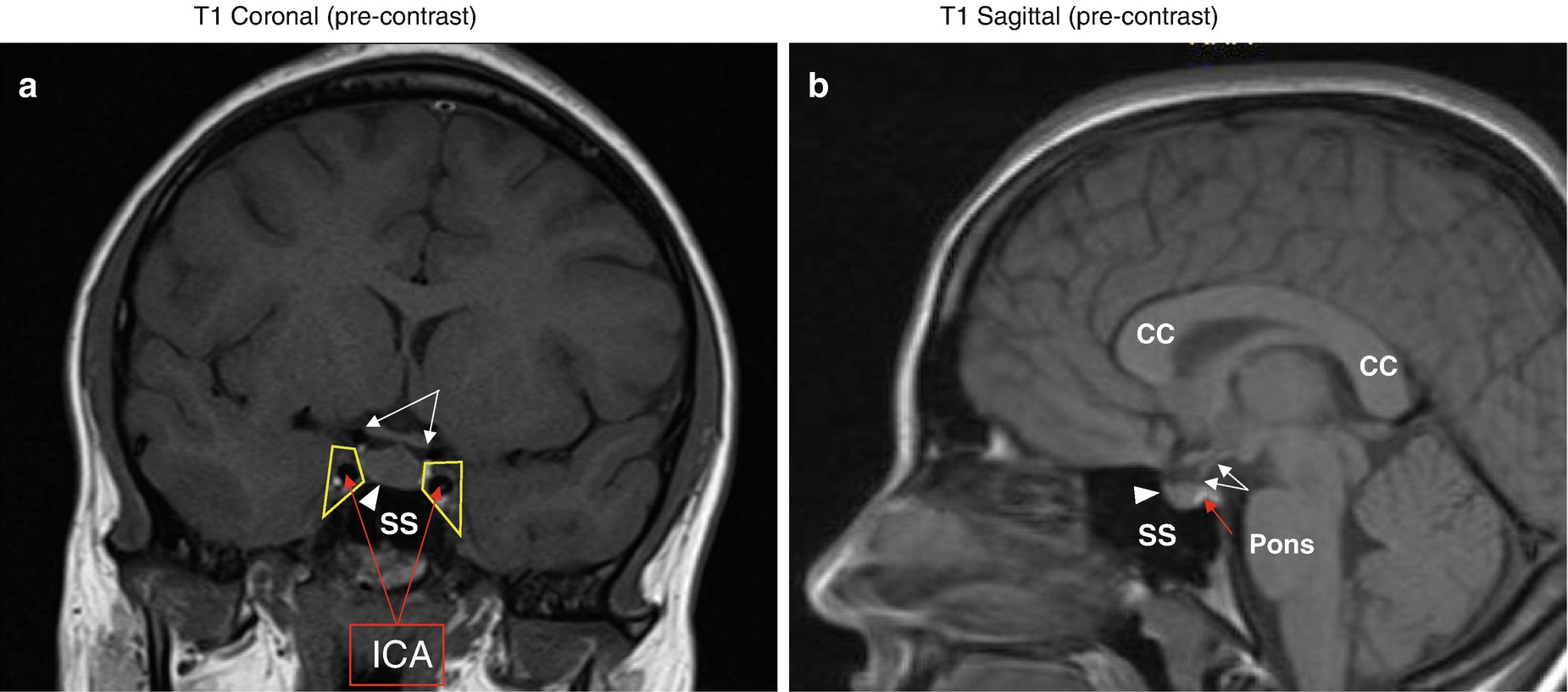
This photo gallery presents the anatomy of pituitary gland by means of MRI T2-weighted coronal views T1-weighted sagittal and coronal views.
Pin On Time To Learn More

Intraoperative Mri Improves Complete Resection Of Pituitary Macroadenoma

Pin On Multiple Sclerosis

Intraoperative Mri Improves Complete Resection Of Pituitary Macroadenoma

Pituitary Center Barrow Neurological Institute
Surgical Treatment Of Pituitary Adenomas Endotext Ncbi Bookshelf
Pituitary Adenoma Johns Hopkins Medicine
Brain With Contrast Showing Normal Enhancement Of The Pituitary Gland Mri At Melbourne Radiology Clinic
Pituitary Gland Mri Scan Protocols Positioning And Positioning Youtube

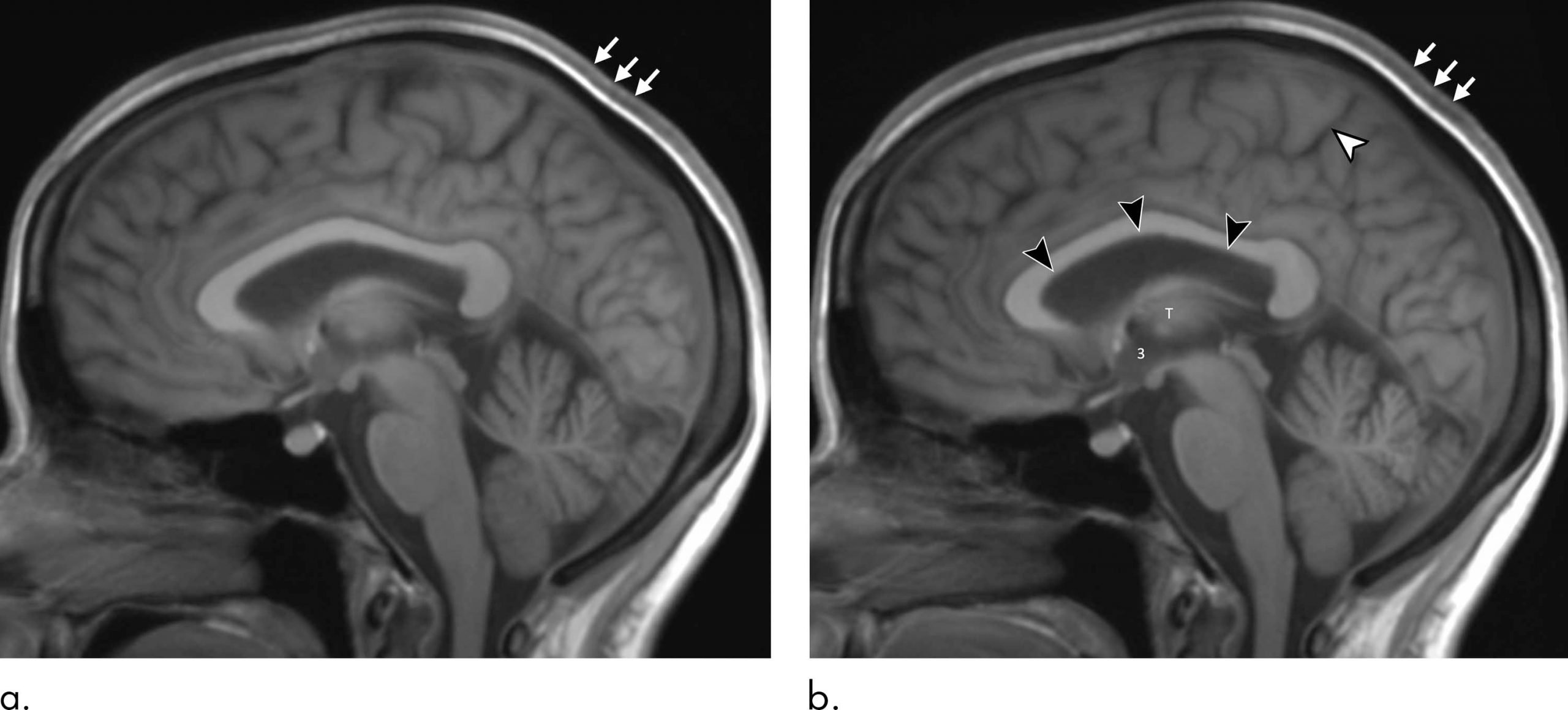
Astronaut Brain Volume Changes And Pituitary Gland Deforms During Long Spaceflights
Pituitary Tumor Adenoma Wills Eye Hospital
Pituitary Adenoma Johns Hopkins Medicine
Pituitary Mri Springerlink
Pituitary Adenoma Johns Hopkins Medicine


Posting Komentar untuk "Pituitary Mri What To Expect"