Shadow On Kidney Ct Scan
Note the pericholecystic fluid but also the fluid collection medial to the posterior liver and lateral to the right kidney as well as free air anterior and medial to the gallbladder. These neuroendocrine tumors are capable of producing and releasing massive amounts of.
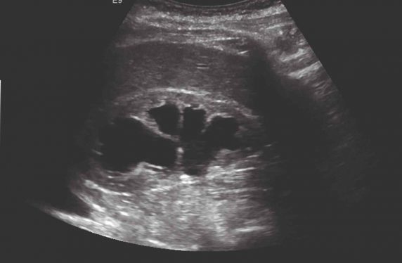
A Curious Case Of A Large Renal Cyst Nephropocus

2016If prediabetes is left untreated 15 to 30 of people with it progress to type 2 diabetes within 5 y American Medical Association and CDC 2015Type 2 diabetes is a major contributor to morbidity mortality and health care.
Shadow on kidney ct scan. Prediabetes is associated with an increased risk of cardiovascular disease coronary heart disease stroke and all-cause mortality Huang et al. As many as one in 11 Americans develop nephrolithiasis and over the past 15 years the prevalence has increased by almost 70 12The number of imaging studies ordered to evaluate for kidney stones is also increasing. A CT scan is usually preferred if its thought the aneurysm has ruptured and theres bleeding on the brain subarachnoid haemorrhage. It can indicate the presence of a tumor but many times these masses are benign noncancerous. There is a tendency for hydrostatic edema to. The diagnosis based on this CT was cardiogenic pulmonary edema.
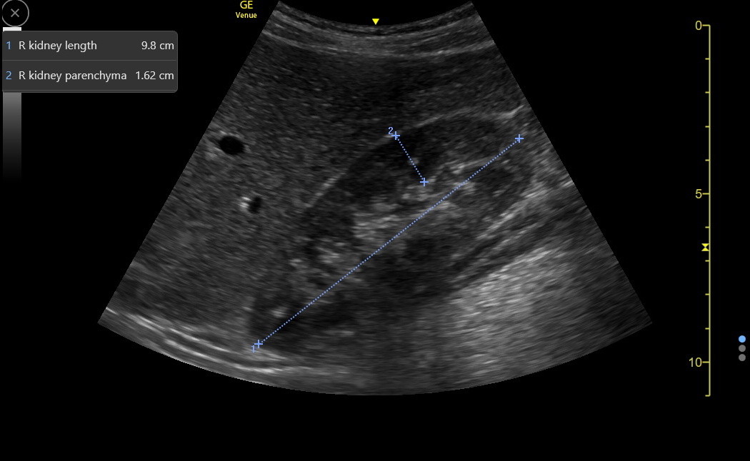
Findings in the Normal Kidney. Computed tomography showed mild splenomegaly and 18F-FDG-PETCT revealed pleural effusion and systemic mild lymphadenopathy. Onset of symptoms may be rapid or more gradual. A tumor derived from embryonal renal cells. Creatinine 118 GFR 49 BUN 17. In some cases a ruptured aneurysm is not picked up by a CT scan.
Metastases of proliferating cells found in the posterior mediastinum. 4k views Answered 2 years ago. The most likely pathophysiology of this childs presentation is. My doctor just started me on Cipro 500 mg 2x daily for 5 days and I am having a CT scan on Monday as he said there were crystals in my urine and indications in the labs that the kidney were not functioning normally. Patient Supine and Upright The AP projection of the abdomen is sometimes called as KUB xray because in this projection the Kidneys Ureters and Bladder are included in the radiographIt is also use as a preliminary evaluation radiograph or Scot films for some of special procedure. When a tumor composed of the same cells as a pheochromocytoma develops outside the adrenal gland it is referred to as a paraganglioma.
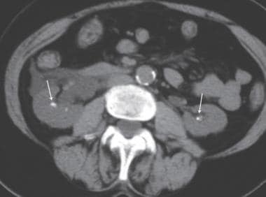
A CT scan is a common non-invasive test that produces a 3-D image of the area scannedIt is used to diagnose conditions and results may be used to determine the best treatment. In the longitudinal scan plane the kidney has the characteristic oval bean-shape. Ct urogram is a better test. CT scan is notable for a unilateral non-calcified mass. Proliferation of catecholamine producing cells. Cardiogenic pulmonary edema generally results in a combination of septal thickening and ground-glass opacity.
Symptoms include abdominal pain abdominal bloating vomiting constipation and bloody stool. Earn real cash back shopping online in-store with Ibotta. I have just recently in the last week had the most terrible nights sleeps. A confirmatory CT scan shown below was requested by the surgical consult. Page Contents1 OVERVIEW2 ORIENTATIONS USED FOR ABDOMINAL X-RAYS3 ANATOMY ON ABDOMINAL X-RAY4 APPROACH GECkoS5 GAS PATTERN INTRALUMINAL6 EXTRALUMINAL GAS7 CALCIFICATIONS8 SOFT TISSUE MASSES OVERVIEW This page is dedicated to providing a guide on the approach to interpreting an abdominal X-ray. Clinicians in a range of medical specialties will encounter patients with kidney stones.
KUB - Abdomen. The 3057 patients in this study had diabetes detected by screening and were randomized to receive either. A volvulus is when a loop of intestine twists around itself and the mesentery that supports it resulting in a bowel obstruction. If they occur near an airway they may cause an obstruction resulting in pneumonia and bronchiectasis. The body casts a shadow on film when it is exposed to the x-ray much like when you hold a flashlight up to your hand and cast a shadow on a wall. Trace the course of the ureter from the pelvis of the kidney along the tips of the lumbar spine transverse processes over the sacroiliac joint down to the ischial spine and medially to the bladder.
From 1992 to 2009 the use of CT for imaging patients with kidney. MENU Early intensive multifactorial blood pressure cholesterol management in patients with type 2 diabetes mellitus was associated with a small nonsignificant reduction in the incidence of cardiovascular disease events and death in a multinational European study. The right kidney is often found more caudally and is slimmer than the left kidney which may have a so-called dromedary hump due to its proximity to the spleen The kidney is surrounded by a capsule separating the kidney from the echogenic perirenal. 8090 of renal tract stones are radio-opaque but will require non-contrast CT or USS to confirm their position in the ureter. This type of scan is also sometimes. They are usually found accidentally when a chest X-ray or chest computed tomography CT scan is done for some other reason.
CT to look at kidney. Ct done to look at kidney to replace the test called an ivp. Before her death seven months later cancer had spread to her kidney liver spine and pubic bone. A CT scan was ordered which showed advanced lung cancer. A CT scan works around this limitation by capturing one narrow slice of your body. A hypoechoic mass is an area on an ultrasound that is more solid than usual tissue.
Pheochromocytoma PHEO or PCC is a rare tumor of the adrenal medulla composed of chromaffin cells also known as pheochromocytes. Register for an Ibotta account today. The mesentery may become so tightly twisted that blood flow to part of the intestine is cut off resulting in. The X-rays wont pass through the contrary so it creates a shadow. All of the tissue that the x-ray passes through overlap on the image making it hard to isolate different elements. A 61-year-old man with a history of hospitalization for acute decompensated heart failure and pulmonary edema was admitted to the intensive cardiac care unit with fatigue shortness of breath and bilateral leg edema.
Hamartomas may occur anywhere on the skin but are especially common on the face lips and neck. Most diabetes experts however agree that a low-carbohydrate high-protein diet is not worth the risk for people with diabetes because they have a high risk of developing kidney disease and a high protein intake can be stressful on the kidneys in those with kidney diseaseDiabetes is the leading cause of kidney failure in the United States. L hilum a trifle pertaining to a hilum. A tumor derived from cells of neural crest origin. This type of scan takes a series of X-rays which are then assembled by a computer into a detailed 3D image. The defendant claimed they were not provided with a full medical history that included a 30-year history of smoking and a mother who died of lung cancer.
Ultrasound vs CT Scans for Kidney Stone Workup A landmark study published in 2014 by the New England Journal of Medicine conducted at 15 various emergency departments evaluated the accuracy of Point of Care Ultrasound POCUS versus computed tomography CT scan as the initial imaging method for patients with suspected kidney stones. The average Ibotta user earns 150 a year on groceries online purchases delivery and more. An MRI is another type of non-invasive test that is used to create a 3-D image of the area scannedIt can be used to determine a diagnosis and a plan of treatment.
Ultrasonography
Renal Abscess Radiology Case Radiopaedia Org Radiology Small Bowel Obstruction Renal

Coronal Reformatted Ct Image After Intravenous Contrast Administration Download Scientific Diagram

Nephrocalcinosis Medullary Sponge Kidney Calculi Stone Calculus Hyperparathyroid Medullary Sponge Kidney Radiology Pet Ct

Kidneys Ureters And Bladder Kub Imaging Practice Essentials Plain Films Of The Abdomen Renal Ultrasonography
Pin On Superficial

A Curious Case Of A Large Renal Cyst Nephropocus

Hypertrophied Column Of Bertin Usually Located At The Junction Of The Upper And Middle Third Of The Kidney And Contain Renal Cortex Th Column Renal Radiology

Xanthogranulomatous Pyelonephritis Radiology Case Radiopaedia Org Radiology Renal Disease Chronic Granulomatous Disease

What To Do With Incidental Kidney Lesions Pulse Today
Pin On Radiography

Longitudinal Scan Of The Right Kidney Showing Echogenic Foci Of Air Download Scientific Diagram
Diagnostic Ct Scans A Ct Scans Obtained In November 2018 Indicated A Download Scientific Diagram
Kidneys And Adrenal Glands


Posting Komentar untuk "Shadow On Kidney Ct Scan"