Radiofrequency Ablation Osteoid Osteoma Recovery
The most common tumor sites were spine n ¼ 22 and lower extremities n ¼ 4. A service of the National Library of Medicine National Institutes of Health.
Osteoid Osteoma Ct Guided Percutaneous Radiofrequency Ablation And Follow Up In 47 Patients Journal Of Vascular And Interventional Radiology
OBQ11234 A 56-year-old male undergoes resection of a mass that was suspected to be a simple lipoma.

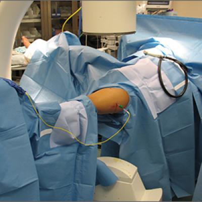
Radiofrequency ablation osteoid osteoma recovery. The benefits of the procedure include having little to no recovery time and nearly immediate pain relief. You also might require more advanced imaging such as MRICT to further evaluate your ankle as it is uncommon for a patient who had osteoid osteoma to. You cant see it but theyre smiling from ear to ear behind those masks. An osteoid osteoma is a benign bone tumor that typically resolves with age or with procedures such as radiofrequency ablation Id be curious to know what you had done for it back in the day. Current trends in treatment of osteoid osteoma with an emphasis on radiofrequency ablation. Osteoid osteomas can affect people of all ages but they occur more frequently in children and young adults.
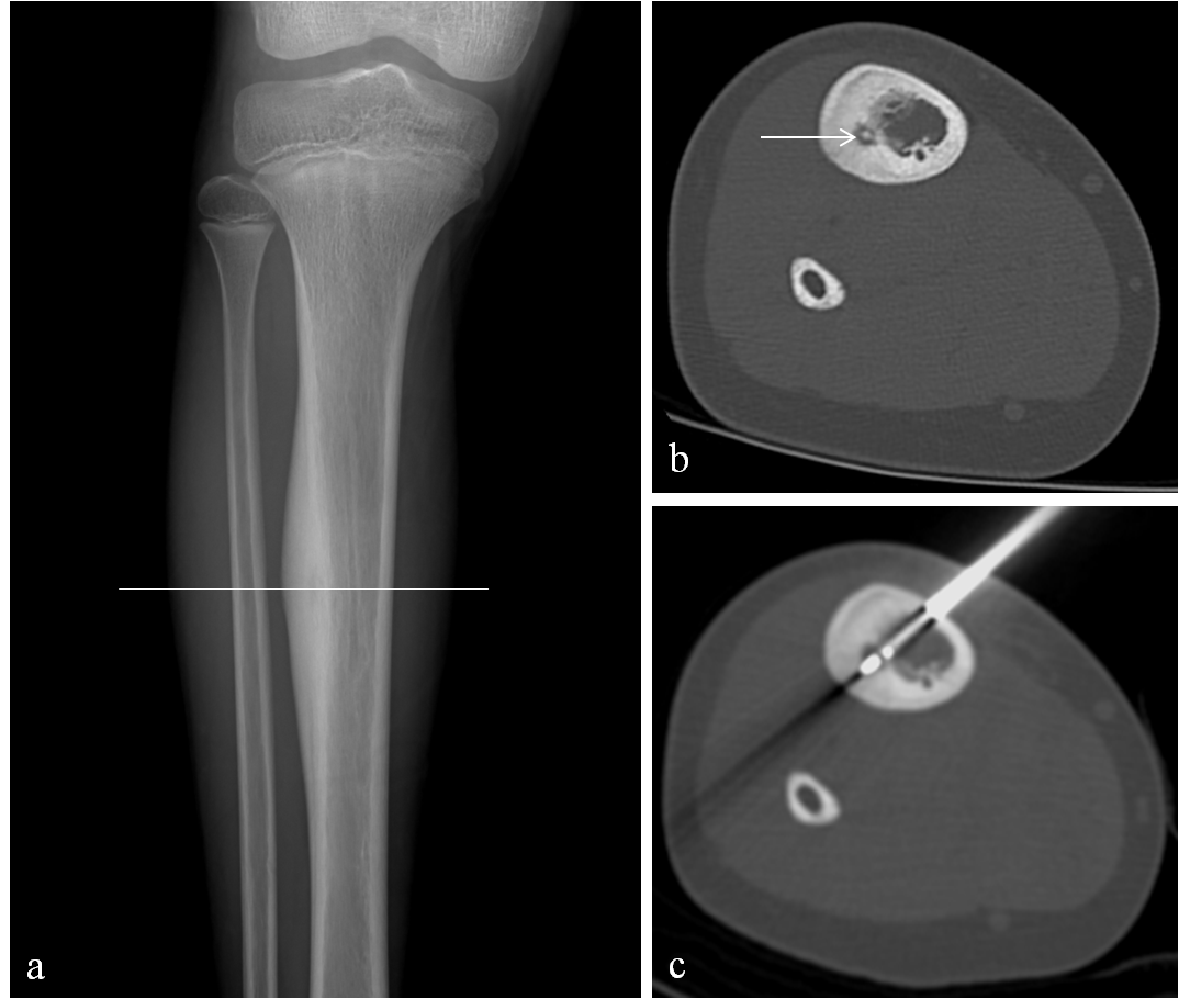
Axial CT image shows a small round lucent lesion with a central calcification at the coracoid base characteristic of an osteoid osteoma. Garge S et al. Osteoid osteoma is a tumor of children and young adults it is very rare in older adults over the age of 50. Crossref Medline Google Scholar. An osteoid osteoma is a benign noncancerous bone tumor that usually develops in the long bones of the body such as the thighbone and shinbone. This procedure uses radiofrequency waves to pulverize the tumor and prevent it from growing back.
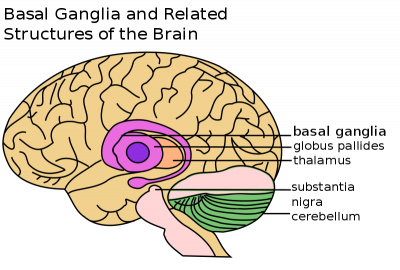
Curr Opin Pediatr 2006. Radiofrequency ablation is a minimally-invasive procedure that uses heat to damage nerves blocking pain signals to the brain. A_ _B_ _C_ _D_ _E_ _F_ _G_ _H_ _I_ _J_ _K_ _L_ _M_ _N_ _O_ _P_ _Q_ _R_ _S_ _T_ _U_ _V_ _W_ _X_ _Y_ _Z_ END A. 69 Cantwell CP Obyrne J Eustace S. HIFU stands for High-Intensity Focused Ultrasound It is also known as MRgFUS MRI-guided focused ultrasound and FUS focused ultrasound surgery. The final pathology came back as a high grade soft tissue sarcoma.
Because our Emory Reproductive Center nurses are the absolute best. As the official journal of the Society of Interventional Radiology JVIR is the peer-reviewed journal of choice for interventional radiologists radiologists cardiologists vascular surgeons neurosurgeons and other clinicians who seek current and reliable information on. HIFU is an innovative non-invasive treatment for a wide range of tumors and diseases. The most common tumor histologies were osteoid osteoma n ¼ 6 venous malformation n ¼ 5 sarcoma n ¼ 5 renal cell carcinoma n ¼ 4 and nonsmall-cell lung cancer n ¼ 3. The management of osteoid osteoma. The mass was contained within his sartorius muscle and shown in Figure A.
HIFU uses an ultrasound transducer similar to the ones used for diagnostic imaging but with much higher energy. Radiofrequency ablation of osteoid osteoma in common and technically challenging locations. JVIR published continuously since 1990 is an international monthly peer-reviewed interventional radiology journal. Interventional radiology IR is a medical subspecialty that performs various minimally-invasive procedures using medical imaging guidance such as x-ray fluoroscopy computed tomography magnetic resonance imaging or ultrasoundIR performs both diagnostic and therapeutic procedures through very small incisions or body orificesDiagnostic IR procedures are those. The patient was successfully treated with CT-guided radiofrequency ablation. Our team of head and neck specialists will create an effective customized treatment plan that is minimally.
This tumor is most frequently found in the legs but may occur also at other bones in nearly any part of the body. Osteoid Osteoma Treated with Minimally Invasive Technique of Radiofrequency Ablation Osteoid osteoma is a benign tumor of the bone. Crossref Medline Google Scholar.
Radiofrequency Ablation For Osteoid Osteoma Southern California Orthopedic Institute

Continued Step By Step Technique Of Radiofrequency Thermal Ablation Download Scientific Diagram

Radiofrequency Ablation Of Osteoid Osteoma With Use Of Intraoperative Three Dimensional Imaging Youtube

Radiofrequency Ablation Of A Spinal Osteoid Osteoma Low Heat Load Technique Journal Of Vascular And Interventional Radiology

Osteoid Osteoma Treated With Radiofrequency Ablation Abstract Europe Pmc

Osteoid Osteoma Hc Chang Orthopaedic Surgery Singapore

Radiofrequency Ablation Of Osteoid Osteoma Initial Results With A Bipolar Ablation Device Journal Of Vascular And Interventional Radiology

Osteoid Osteoma Treatment Stanford Health Care

Bone Ablation Using Image Guidance

Step By Step Technique Of Radiofrequency Thermal Ablation Of Osteoid Download Scientific Diagram

Osteoid Osteoma Of The Talus Treated With Laser Ablation A Sagittal Download Scientific Diagram

Intraoperative Radiofrequency Ablation For Osteoid Osteoma Mdedge Surgery
Oo Ablation
Birpublications Org


Posting Komentar untuk "Radiofrequency Ablation Osteoid Osteoma Recovery"